Framed Print > Animals > Mammals > Muridae > Water Mouse
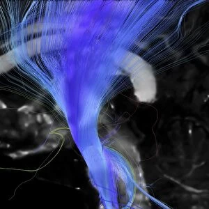
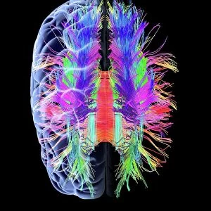
Framed Print : Brain fibres, tract density imaging C017 / 7039


![]()
Framed Photos from Science Photo Library
Brain fibres, tract density imaging C017 / 7039
Brain fibres, tract density imaging. Axial scan of the brain showing the density of nerve pathways (tracts) in the brain. The image was obtained using 3D diffusion tensor imaging (DTI) magnetic resonance imaging (MRI) scans. The fibres are locally coloured red-green-blue if they are orientated in x-y-z alignment (left-right, posterior-anterior, inferior-superior). Diffusion tensor imaging measures the direction of water diffusion, which in the brain reveals the orientation of nerve fibres. The technique is also known as tractography, producing a tractogram
Science Photo Library features Science and Medical images including photos and illustrations
Media ID 9340949
© SHERBROOKE CONNECTIVITY IMAGING LAB/SCIENCE PHOTO LIBRARY
Brain Imaging Brain Scan Central Nervous System Cerebral Cerebrum Diffusion Tensor Imaging Dti Scan Fiber Fibers Fibre Fibres Imaging Technique Magnetic Resonance Imaging Mri Scan Mri Scanner Nerve Nerve Fibre Nerves Neural Pathway Neural Tract Paths Pathway Pathways Structural Tractogram Tractography White Matter Brain Neurological Neurology
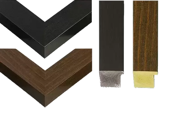
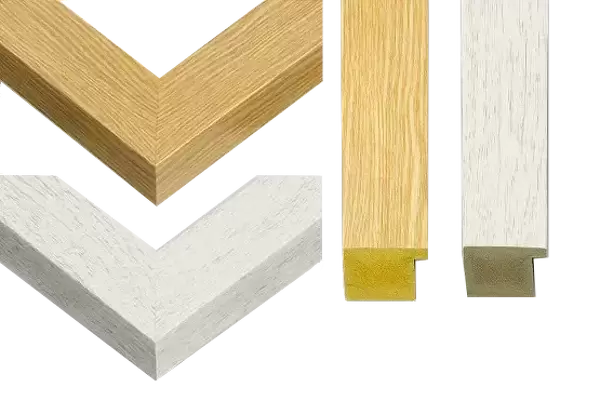
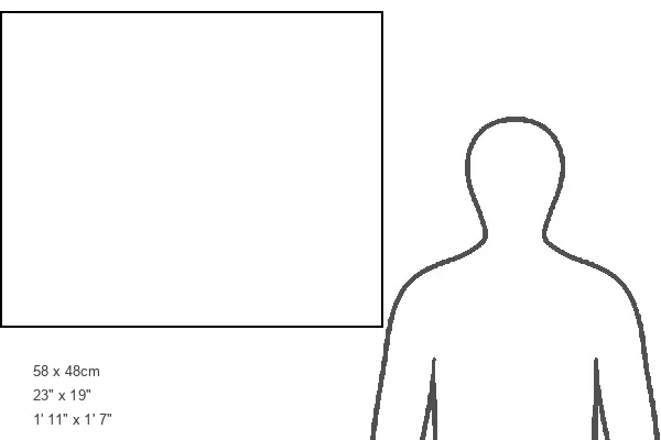
22"x18" (58x48cm) Modern Frame
Discover the intricacies of the human brain with our Framed Prints from Media Storehouse, featuring the captivating image "Brain Fibres, Tract Density Imaging C017 / 7039" by SHERBROOKE CONNECTIVITY IMAGING LAB/SCIENCE PHOTO LIBRARY. This image showcases the brain's complex nerve pathways, or tracts, in stunning detail. Obtained using advanced 3D diffusion tensor imaging (DTI), each fiber is vividly depicted, providing an unparalleled perspective into the intricate neural connections that make up our most complex organ. Elevate your home or office décor while celebrating the wonders of neuroscience with this thought-provoking and visually stunning addition to your space.
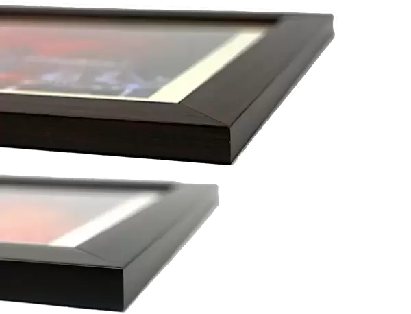
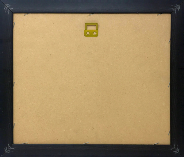
Wood effect frame, card mounted, 16x12 archival quality photo print. Overall outside dimensions 22x18 inches (58x48cm). Environmentally and ozone friendly, 40mm wide x 15mm Polycore® moulding has the look of real wood, is durable and light and easy to hang. Biodegradable and made with non-chlorinated gases (no toxic fumes) it is efficient; producing 100 tons of polystyrene can save 300 tons of trees! Prints are glazed with lightweight, shatterproof, optical clarity acrylic (providing the same general protection from the environment as glass). The back is stapled hardboard with a sawtooth hanger attached. Note: To minimise original artwork cropping, for optimum layout, and to ensure print is secure, the visible print may be marginally smaller
Contemporary Framed and Mounted Prints - Professionally Made and Ready to Hang
Estimated Image Size (if not cropped) is 39.6cm x 27cm (15.6" x 10.6")
Estimated Product Size is 57.9cm x 47.8cm (22.8" x 18.8")
These are individually made so all sizes are approximate
Artwork printed orientated as per the preview above, with landscape (horizontal) orientation to match the source image.
FEATURES IN THESE COLLECTIONS
> Animals
> Mammals
> Muridae
> Water Mouse
EDITORS COMMENTS
This print showcases the intricate network of brain fibres, providing a glimpse into the complex pathways that make up our central nervous system. With a black background serving as a canvas, the image highlights the density and arrangement of nerve tracts within the brain using vibrant red, green, and blue colors. Each color represents a specific orientation in three-dimensional space: left-right (x-axis), posterior-anterior (y-axis), and inferior-superior (z-axis). Obtained through advanced 3D diffusion tensor imaging (DTI) magnetic resonance imaging (MRI) scans, this technique called tractography allows researchers to visualize and map out neural connections within the brain. By measuring water diffusion direction, DTI reveals the orientation of nerve fibres throughout various regions. The photograph not only captures the beauty of these intricate structures but also holds significant implications for fields such as biology, medicine, neurology, and anatomy. It provides valuable insights into normal brain function and structure while offering potential diagnostic applications for understanding neurological disorders. As we delve deeper into understanding how our brains work on both anatomical and functional levels, images like this serve as powerful tools for unraveling mysteries hidden within our most vital organ – paving the way for advancements in medical research and ultimately improving human health.
MADE IN THE UK
Safe Shipping with 30 Day Money Back Guarantee
FREE PERSONALISATION*
We are proud to offer a range of customisation features including Personalised Captions, Color Filters and Picture Zoom Tools
SECURE PAYMENTS
We happily accept a wide range of payment options so you can pay for the things you need in the way that is most convenient for you
* Options may vary by product and licensing agreement. Zoomed Pictures can be adjusted in the Basket.