
Nerve Fibre Collection
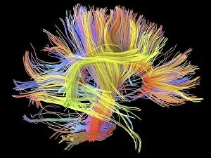
"Nerve Fibres: Unveiling the Intricacies of Brain Connectivity" Delving into the depths of our complex neural network


All Professionally Made to Order for Quick Shipping
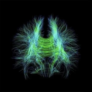
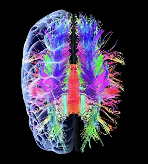
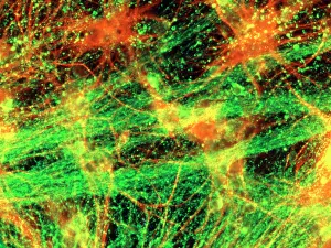
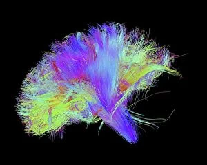
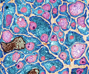
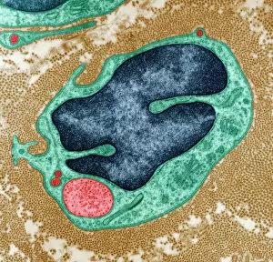
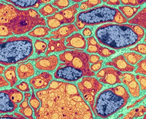
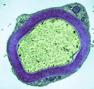
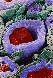
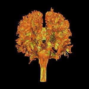
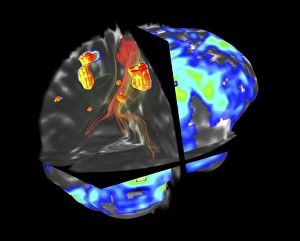
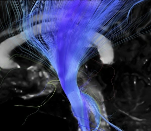
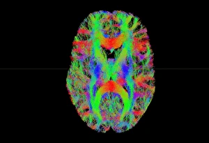
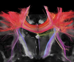
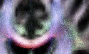
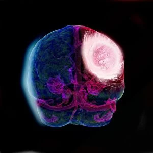
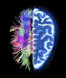
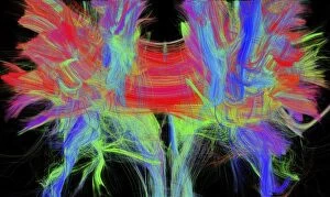
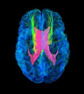
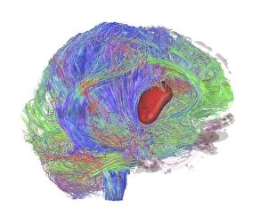
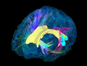
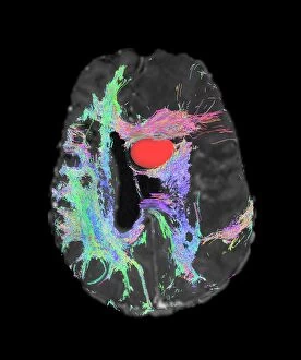
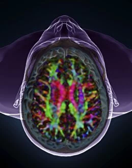
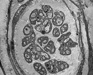
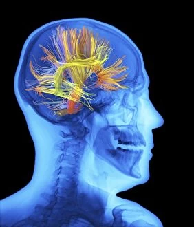
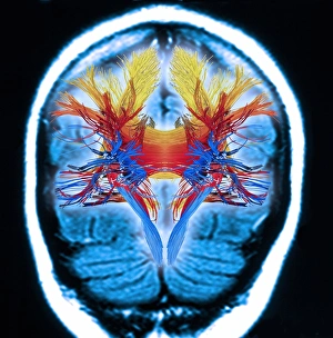
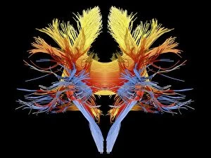
"Nerve Fibres: Unveiling the Intricacies of Brain Connectivity" Delving into the depths of our complex neural network, nerve fibres serve as the intricate highways that facilitate communication within our brain. Through cutting-edge technology such as DTI MRI scans and immunofluorescent LM, scientists have begun unraveling the mysteries hidden within these delicate structures. The mesmerizing artwork depicting white matter fibres intertwined with the brain showcases their vital role in transmitting information across different regions. In C017 / 7099 and C017 / 7035 scans, we witness a captivating glimpse into this web-like connectivity, highlighting how these fibres enable seamless coordination between various cognitive processes. Immersing ourselves further into this microscopic realm, TEM images reveal regenerating nerve cells undergoing a remarkable transformation. Witnessing their resilience and ability to heal inspires awe at nature's incredible capacity for regeneration. Myelination emerges as another fascinating aspect captured by TEM imagery. The protective sheath surrounding nerve fibres ensures efficient transmission of electrical impulses, enhancing signal conduction speed and overall neural efficiency. As we explore diverse subjects like bee anatomy or artistic representations of white matter fibres in C014 / 5666 and C014 / 5668 artworks, we gain a deeper appreciation for both scientific inquiry and creative expression intertwining to shed light on these enigmatic structures. Through continued research on nerve fibre function and connectivity patterns, scientists strive to unlock new insights into neurological disorders such as Alzheimer's disease or multiple sclerosis. These investigations hold immense promise for developing targeted therapies aimed at preserving or restoring optimal brain function. Intricate yet resilient, nerve fibres stand as testaments to the complexity of human cognition. As science advances hand-in-hand with artistry in capturing their essence, humanity moves closer towards understanding one of its most profound creations – the human mind itself.