Fraud Blocker Skip to main content

Neurology Collection

Neurology, the fascinating study of the nervous system, unravels the intricate workings of our brain and its various components

Background imageNeurology Collection: Motor homunculus model

Motor homunculus model. Parts of the body are sized according to how much space the brain gives to controlling the movement of that part of the body

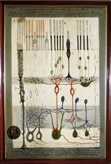
Background imageNeurology Collection: Histological Diagram of a Mammalian Retina

Histological Diagram of a Mammalian Retina
RAMON Y CAJAL, Santiago (1852-1934). Spanish doctor and histologist, Nobel Prize in 1906. Histological diagram of a mammalian retina. Original drawing by Ramon y Cajal

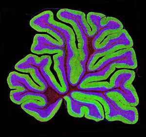
Background imageNeurology Collection: Cerebellum tissue, light micrograph

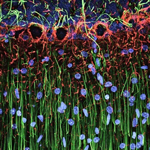
Cerebellum tissue, light micrograph
Cerebellum tissue. Confocal light micrograph of a section through the cerebellum of the brain. Purkinje cells, a type of neuron (nerve cell), are red

Background imageNeurology Collection: Brain pathways

Brain pathways

Background imageNeurology Collection: Brain fibres, DTI MRI scan C017 / 7099

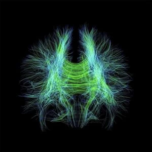
Brain fibres, DTI MRI scan C017 / 7099
Brain fibres. 3D diffusion tensor imaging (DTI) magnetic resonance imaging (MRI) scan of nerve pathways in the brain. The pathways are highlighted in green and blue. The brain is seen from the front

Background imageNeurology Collection: Anatomy of human brain, inferior view

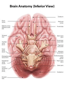
Anatomy of human brain, inferior view

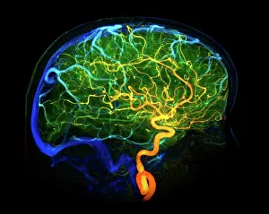
Background imageNeurology Collection: Brain blood vessels, 3D angiogram C007 / 1981

Brain blood vessels, 3D angiogram C007 / 1981
Brain blood vessels. Coloured 3D lateral angiogram (blood vessel X-ray) of the blood vessels in the brain of a healthy 32-year-old

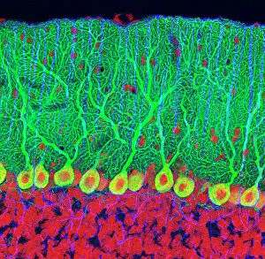
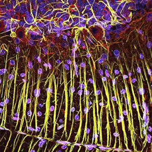
Background imageNeurology Collection: Purkinje nerve cells in the cerebellum

Purkinje nerve cells in the cerebellum
Purkinje cells in the cerebellum. Fluorescent light micrograph of Purkinje cells (green) in the cerebellum of the brain. Purkinje nerve cells have a flask-like body from which numerous highly

Background imageNeurology Collection: Medulla oblongata in the brain, artwork

Medulla oblongata in the brain, artwork
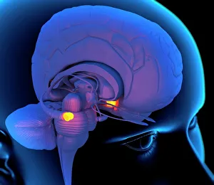
Hypothalamus in the brain. Computer artwork of a persons head showing the brain inside. The highlighted area shows the hypothalamus

Background imageNeurology Collection: Brain fibres, DTI MRI scan C017 / 7035

Brain fibres, DTI MRI scan C017 / 7035
Brain fibres. 3D diffusion tensor imaging (DTI) magnetic resonance imaging (MRI) scan of nerve pathways in the brain. The pathways are highlighted in blue

Background imageNeurology Collection: Motor and sensory homunculi

Motor and sensory homunculi
Sensory and motor homunculi. Computer artwork of a motor homunculus (left) and a sensory homunculus (right). Parts of the body are sized according to how much space the brain gives to processing

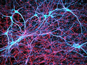
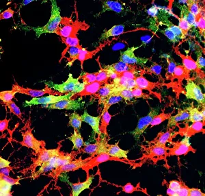
Background imageNeurology Collection: Nerve and glial cells, light micrograph

Nerve and glial cells, light micrograph
Nerve and glial cells, fluorescence light micrograph. These are neural stem cells that have differentiated into neurons (nerve cells, blue) and glial cells (support cells, red)

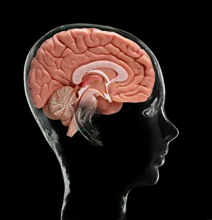
Background imageNeurology Collection: Human brain, model

Human brain, model. Composite image of a sectioned model human brain inside a glass head. This view shows some of the brains major structures, including: the cerebral cortex (folded areas of tissue)

Background imageNeurology Collection: Caffeine crystals, light micrograph

Caffeine crystals, light micrograph
Caffeine crystals. Polarised light micrograph of crystals of caffeine (1, 3, 7-trimethylxanthine). Caffeine stimulates the central nervous system increasing alertness and deferring fatigue

Background imageNeurology Collection: Hippocampus brain tissue

Hippocampus brain tissue
Hippocampus tissue. Light micrograph of a sagittal (side view) section through the hippocampus of the brain showing the nerve cells within it

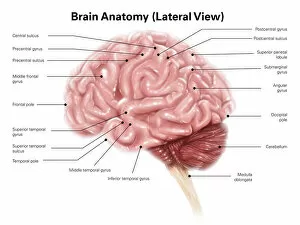
Background imageNeurology Collection: Human brain anatomy, lateral view

Human brain anatomy, lateral view

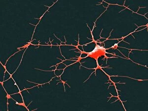
Background imageNeurology Collection: Nerve cell, SEM

Nerve cell, SEM
Nerve cell. Coloured scanning electron micrograph (SEM) of a nerve cell (neuron). Neurons are responsible for passing information around the central nervous system (CNS)

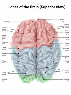
Background imageNeurology Collection: Superior view of human brain with colored lobes and labels

Superior view of human brain with colored lobes and labels

Background imageNeurology Collection: RAMON Y CAJAL, Santiago (1852-1934). Spanish

RAMON Y CAJAL, Santiago (1852-1934). Spanish

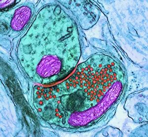
Background imageNeurology Collection: Synapse nerve junction, TEM

Synapse nerve junction, TEM
Synapse. Coloured transmission electron micrograph (TEM) of a synapse, a junction between two nerve cells, in the brain. At a synapse an electrical signal is transmitted from one cell to the next in

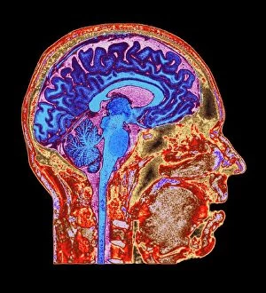
Background imageNeurology Collection: Brain anatomy, MRI scan

Brain anatomy, MRI scan
Brain anatomy. Coloured magnetic resonance imaging (MRI) scan of the human head from the side. The sagittal scan has halved the brain, revealing the internal anatomy

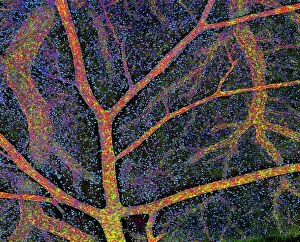
Background imageNeurology Collection: Brain tissue blood supply

Brain tissue blood supply. Light micrograph of a section through cortex tissue from a brain, showing the blood vessels (branching) that supply it

Background imageNeurology Collection: Medulla oblongata in the brain, artwork

Medulla oblongata in the brain, artwork
Hypothalamus in the brain. Computer artwork of a persons head showing the left hemisphere of the brain inside. The highlighted area shows the hypothalamus

Background imageNeurology Collection: Nervous system

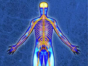
Nervous system. Computer artwork of the principle nerve pathways of the human body. The brain (top centre) and the spinal cord (down centre) comprise the central nervous system (CNS)

Background imageNeurology Collection: Nerve cell

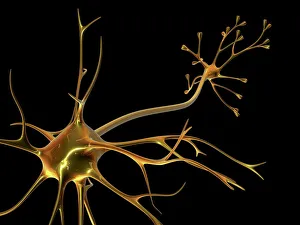
Nerve cell. Computer artwork of a nerve cell, also called a neuron. Neurons are responsible for passing information around the central nervous system (CNS) and from the CNS to the rest of the body

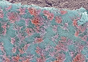
Background imageNeurology Collection: Brain surface, SEM

Brain surface, SEM
Brain surface. Coloured scanning electron micrograph (SEM) of the surface of a brain ventricle. Ventricles are cavities within the brain that are continuous with the central canal of the spinal cord

Background imageNeurology Collection: Basal ganglia, artwork

Basal ganglia, artwork
Basal ganglia. Computer artwork of the human brain, showing some of the structures that make up the basal ganglia (basal nuclei, pink)

Background imageNeurology Collection: Glial stem cell culture, light micrograph

Glial stem cell culture, light micrograph
Glial stem cell culture. Fluorescent light micrograph of glial stem cells producing the protein NG2 (red) as they mature. These stem cells can differentiate into several types of glial cells

Background imageNeurology Collection: Cerebral cortex nerve cells

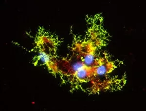
Cerebral cortex nerve cells. Confocal light micrograph of neurons (nerve cells, red) and glial cells (support cells, gold) from the cerebral cortex
Background imageNeurology Collection: Purkinje nerve cells in the cerebellum

Purkinje nerve cells in the cerebellum
Purkinje cells in the cerebellum. Fluorescent light micrograph of Purkinje cells (green) in the cerebellum of the brain. Purkinje nerve cells have a flask-like body from which numerous highly

Background imageNeurology Collection: Regenerating nerve cell, TEM

Regenerating nerve cell, TEM
Regenerating nerve cell. Coloured transmission electron micrograph (TEM) of a section through a nerve axon (blue) regenerating within a Schwann cell (light brown)

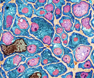
Background imageNeurology Collection: Myelination of nerve fibres, TEM

Myelination of nerve fibres, TEM
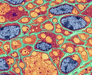
Myelination of nerve fibres. Coloured transmission electron micrograph (TEM) of Schwann cells (blue, with brown nuclei) insulating nerve fibres (axons, pink) with a myelin sheath

Background imageNeurology Collection: Cerebellum structure, light micrograph

Cerebellum structure, light micrograph
Cerebellum structure. Fluorescent light micrograph of a section through the cerebellum of the brain. The cerebellum comprises three main layers

Background imageNeurology Collection: Brain protein research

Brain protein research. Computer artwork of a brain and coloured dots from a protein microarray. Protein microarrays can be used to follow protein interactions

Background imageNeurology Collection: Neural stem cell culture

Neural stem cell culture. Fluorescent light micrograph of a group of neural stem cells (neurosphere) in culture. Neural stem cells are able to differentiate into neurons (nerve cells)

Background imageNeurology Collection: Regenerating nerve cell, TEM

Regenerating nerve cell, TEM
Regenerating nerve cell. Coloured transmission electron micrograph (TEM) of a section through a nerve axon (yellow) regenerating within a Schwann cell (blue). The Schwann cells nucleus is black

Background imageNeurology Collection: Motor neurons, light micrograph

Motor neurons, light micrograph. Motor neurons are responsible for passing information around the central nervous system (CNS) and from the CNS to the rest of the body

Background imageNeurology Collection: Cerebellum tissue, light micrograph

Cerebellum tissue, light micrograph
Cerebellum tissue. Confocal light micrograph of a section through the cerebellum of the brain showing two types of glial cells (support cells); astrocytes (star-shaped)

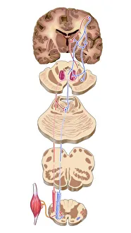
Background imageNeurology Collection: Brain motor cortex pathways, artwork C016 / 6532

Brain motor cortex pathways, artwork C016 / 6532
Brain motor cortex pathways. Artwork of a sectioned human brain, brainstem and spinal cord, showing neural pathways (red and blue) from the motor cortex of the brain

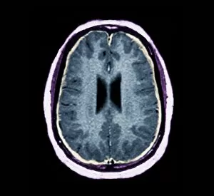
Background imageNeurology Collection: Bacterial meningitis, MRI scan

Bacterial meningitis, MRI scan
Bacterial meningitis. Coloured magnetic resonance imaging (MRI) scan of an axial section through the brain of a 16-year-old patient

Background imageNeurology Collection: Brain neuron

Brain neuron. Computer reconstruction of a medium spiny neuron from the basal ganglia of the brain. Neurons (nerve cells) are responsible for passing information around the central nervous system

Background imageNeurology Collection: Cerebellum tissue, light micrograph

Cerebellum tissue, light micrograph
Cerebellum tissue. Confocal light micrograph of a section through the cerebellum of the brain. Purkinje cells, a type of neuron (nerve cell), are red

Background imageNeurology Collection: Normal human brain, MRI scan C016 / 8845

Normal human brain, MRI scan C016 / 8845
Brain. Coloured magnetic resonance imaging (MRI) scan of a sagittal section through a patients head showing a healthy human brain

Background imageNeurology Collection: Phantom pain after amputation, artwork

Phantom pain after amputation, artwork
Phantom pain after amputation. Artwork of a man experiencing phantom pain (red flash) from his amputated hand (left). This is caused by stimuli to the motor and pre-motor cortical areas of his brain

Background imageNeurology Collection: Nerve cell, TEM

Nerve cell, TEM
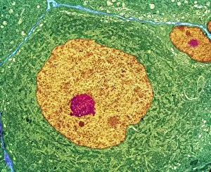
Nerve cell. Coloured transmission electron micrograph (TEM) of a nerve cell body in cross- section. The cell has a large nucleus (yellow) and inner nucleolus (red)

Background imageNeurology Collection: Oligodendrocyte nerve cells

Oligodendrocyte nerve cells. Fluorescent light micrograph of human oligodendrocyte nerve cells. Cell nuclei, which contain the cells genetic information, have been dyed blue

Background imageNeurology Collection: Nerve cells, abstract artwork

Nerve cells, abstract artwork
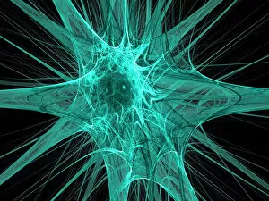
Nerve cells. Abstract computer artwork of nerve cells, or neurons. Neurons are responsible for passing information around the central nervous system (CNS) and from the CNS to the rest of the body

Background imageNeurology Collection: Myelination of nerve fibres, TEM

Myelination of nerve fibres, TEM
Myelination of nerve fibres. Coloured transmission electron micrograph (TEM) of Schwann cells (red, with blue nuclei) insulating nerve fibres (axons, orange) with a myelin sheath


All Professionally Made to Order for Quick Shipping

Neurology, the fascinating study of the nervous system, unravels the intricate workings of our brain and its various components. From the Motor homunculus model that depicts how different body parts are represented in our motor cortex to the Histological Diagram of a Mammalian Retina revealing the complexity of our visual processing, every aspect is awe-inspiring. The Cerebellum tissue, as seen through a light micrograph, showcases its unique structure responsible for coordinating movement and balance. Meanwhile, an inferior view of the Anatomy of human brain highlights its remarkable organization and interconnectedness. Through Brain fibres captured by DTI MRI scans like C017 / 7099 or C017 / 7035, we gain insights into neural pathways that facilitate communication between different regions. These pathways form a complex network crucial for transmitting information throughout our brain. Exploring Brain blood vessels using a 3D angiogram (C007 / 1981) reveals their vital role in supplying oxygen and nutrients to sustain neuronal function. Additionally, understanding Motor and sensory homunculi helps us comprehend how specific areas within our brains control different bodily functions. Delving deeper into neurology brings us face-to-face with Medulla oblongata artwork depicting this critical region involved in regulating essential autonomic functions such as breathing and heart rate. Furthermore, observing Nerve and glial cells under a light microscope illustrates their diverse roles in supporting neurons' health and functionality. Finally, examining Synapse nerve junctions through TEM unveils these microscopic structures where electrical signals are transmitted from one neuron to another—a fundamental process underlying all brain activity. In essence, neurology takes us on an incredible journey deep within ourselves—unveiling mysteries at both macroscopic levels like anatomical structures or microscopically exploring cellular intricacies, and is through this exploration that we gain profound knowledge about how our brains work—the very essence of who we are as individuals.