
Dti Scan Collection
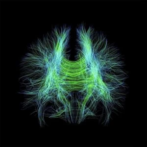
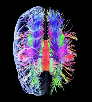
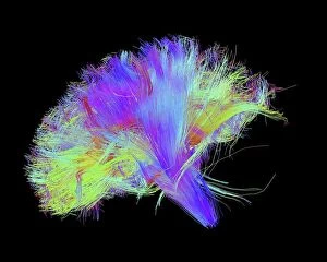
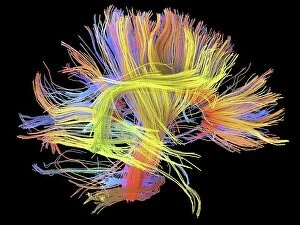
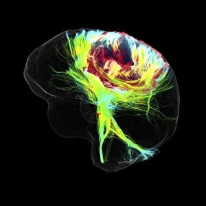
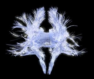
"Unraveling the Intricacies of Brain Fibres: A Journey through DTI Scans" Delve into the fascinating world of brain fibres with DTI MRI scans


All Professionally Made to Order for Quick Shipping
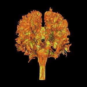
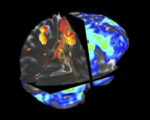
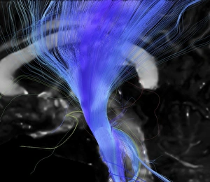
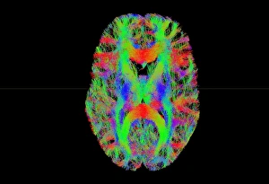
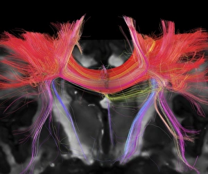
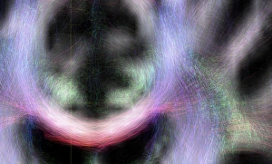
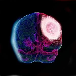
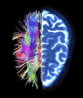
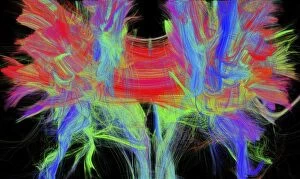
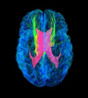
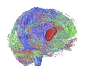
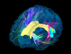
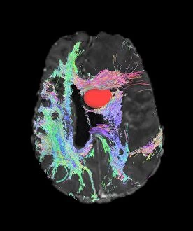
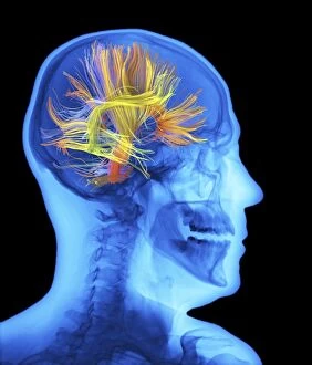
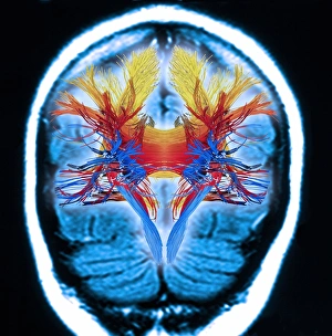
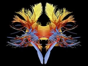
"Unraveling the Intricacies of Brain Fibres: A Journey through DTI Scans" Delve into the fascinating world of brain fibres with DTI MRI scans. These advanced imaging techniques, such as C017 / 7099 and C017 / 7035, allow us to explore the intricate network of white matter fibres in the human brain. Step into a realm where art meets science with artwork C015 / 1930, showcasing the beauty and complexity of these brain fibres. Marvel at their delicate structure and how they connect different regions within our minds. Witness the stunning visuals captured by scans like C014 / 5666, C014 / 5668, and C014 / 5667 that reveal the mesmerizing patterns formed by white matter fibres. Each scan provides a unique glimpse into this essential component of our brains. Explore how DTI scans shed light on various neurological conditions. Discover how fMRI and tractography help identify brain tumours (C017 / 7102) or examine specific pathways like corticospinal tracts (C017 / 7046). With tract density imaging (C017/7039), we can further understand the distribution and connectivity of these vital fibres. Immerse yourself in this captivating journey through brains' white matter using DTI MRI scans. Witness firsthand how these cutting-edge technologies enable us to visualize complex structures that were once hidden from view. Unlocking new insights about brain function and pathology is made possible by models like those seen in C017/7060 which provide valuable information about tumour growth using DTI modelling techniques. Intriguingly beautiful yet scientifically significant, DTI scans offer an unparalleled window into understanding brain fibre networks.