
Fibres Collection
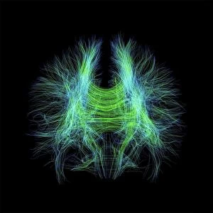
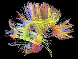
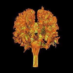
"Fibres: Unraveling the Intricacies of the Human Brain and Beyond" Exploring the intricate network of brain fibres through DTI MRI scans (C017 / 7099


All Professionally Made to Order for Quick Shipping
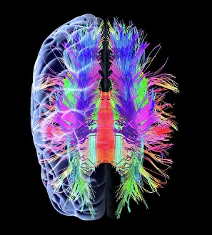
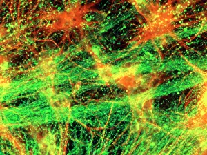
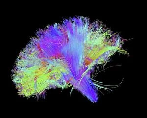
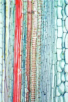
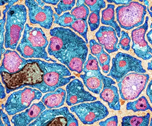
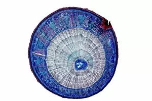
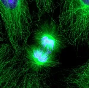
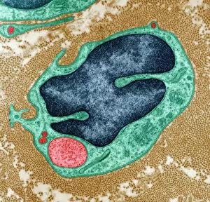
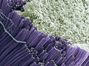
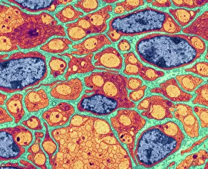
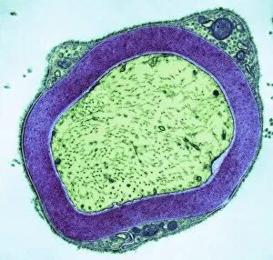
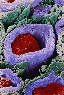
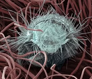
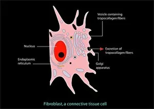
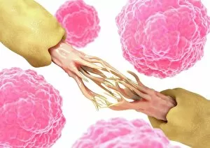
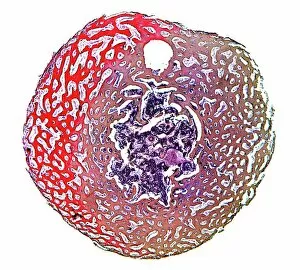
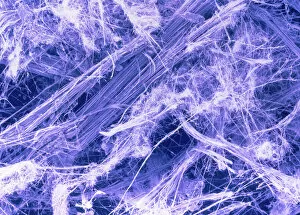
"Fibres: Unraveling the Intricacies of the Human Brain and Beyond" Exploring the intricate network of brain fibres through DTI MRI scans (C017 / 7099). The mesmerizing artwork depicting white matter fibres and their connection to the brain (C015 / 1930). Delving deeper into understanding brain fibres with DTI MRI scans (C017 / 7035). Illuminating neurons and astrocytes through immunofluorescent LM, revealing the complexity of brain fibres. Peering into the white matter fibres of the human brain, unlocking its mysteries (C014 / 5666). Witnessing nerve cells regenerate under a TEM microscope, showcasing nature's resilience. Marveling at the intricate structure of a lime tree stem captured in a light micrograph. Discovering beauty in unexpected places - castor oil stem captured in a mesmerizing light micrograph. Unveiling myelination's role in enhancing nerve fibre function through TEM imaging. Witnessing cell division unfold under fluorescent micrographs, highlighting life's continuous renewal process. Celebrating nature's ability to heal as we observe regenerating nerve cells under TEM microscopy once again. Exploring further into white matter fibres within our complex human brains (C014/5668). In this captivating journey, we unravel the wonders – from delicate neural connections within our brains to resilient plant stems that provide sustenance for life itself.