Fraud Blocker Skip to main content

Fibres Collection (page 6)

"Fibres: Unraveling the Intricacies of the Human Brain and Beyond" Exploring the intricate network of brain fibres through DTI MRI scans (C017 / 7099

Background imageFibres Collection: Ancient Peruvian fabric C016 / 2368

Ancient Peruvian fabric C016 / 2368
Ancient Peruvian fabric. Section of fabric dating from the Nazca-Huari Fusion Epoch (800AD-1300AD). This textile is decorated with stepped motifs and concentric interlaced rhombuses
Background imageFibres Collection: Fibre optics factory C013 / 9199

Fibre optics factory C013 / 9199
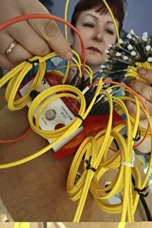
Fibre optics factory. Worker with complete optical patch cables at a factory that produces fibre optic equipment. Optical fibres are made from flexible glass that has a high refractive index

Background imageFibres Collection: Fibre optics factory C013 / 9200

Fibre optics factory C013 / 9200
Fibre optics factory. Worker with complete optical patch cables at a factory that produces fibre optic equipment. Optical fibres are made from flexible glass that has a high refractive index

Background imageFibres Collection: Fibre optics factory C013 / 9193

Fibre optics factory C013 / 9193
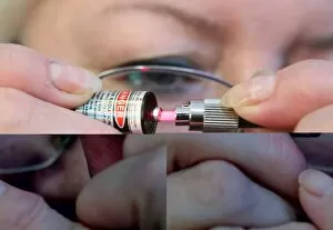
Fibre optics factory. Worker using a laser to check the optical contact of an optical cable at a factory that produces fibre optic equipment

Background imageFibres Collection: Myelinated nerves, SEM C013 / 7142

Myelinated nerves, SEM C013 / 7142
Myelinated nerves. Coloured scanning electron micrograph (SEM) of a section through the sciatic nerve, showing the myelinated nerve fibres (axons)

Background imageFibres Collection: Myelinated nerves, SEM C013 / 7141

Myelinated nerves, SEM C013 / 7141
Myelinated nerves. Coloured scanning electron micrograph (SEM) of a section through the sciatic nerve, showing the myelinated nerve fibres (axons)

Background imageFibres Collection: Myelinated nerves, SEM C013 / 7138

Myelinated nerves, SEM C013 / 7138
Myelinated nerves. Coloured scanning electron micrograph (SEM) of a section through a myelinated nerve fibre (axon, beige, centre) from the sciatic nerve

Background imageFibres Collection: Synthetic sponge, SEM

Synthetic sponge, SEM
Synthetic sponge. Coloured scanning electron micrograph (SEM) showing the branching structure of a synthetic sponge. This structure is based on that of natural sponges

Background imageFibres Collection: Natural sponge, SEM

Natural sponge, SEM
Natural sponge. Coloured scanning electron micrograph (SEM) showing the branching structure of the body of a natural sponge (phylum porifera)

Background imageFibres Collection: Balsa wood structure, SEM

Balsa wood structure, SEM
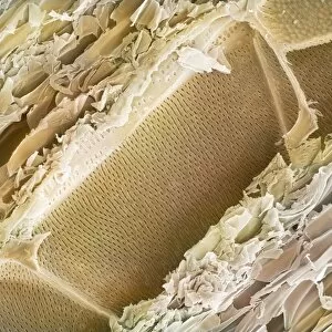
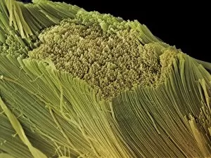
Balsa wood structure, coloured scanning electron micrograph (SEM). This wood is from the balsa tree (Ochroma pyramidale). This is a longitudinal section through the wood

Background imageFibres Collection: Kitchen paper towel, SEM

Kitchen paper towel, SEM
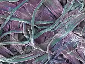
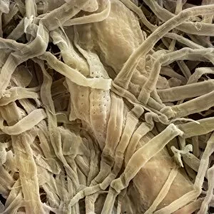
Kitchen paper towel fibres. Coloured scanning electron micrograph (SEM) of fibres from a sheet of kitchen towel. Paper mostly consists of a mesh of softened and pulped wood fibres

Background imageFibres Collection: Paper handkerchief, SEM

Paper handkerchief, SEM
Paper handkerchief fibres. Coloured scanning electron micrograph (SEM) of a paper hankerchief, or tissue, which consists mostly of softened and pulped wood fibres

Background imageFibres Collection: Terylene Hat

Terylene Hat
A trendy Terylene pork pie hat! Date: 1960s

Background imageFibres Collection: Fake Fur Hat

Fake Fur Hat
A gentleman sporting a jaunty fake fur hat! Date: 1960s

Background imageFibres Collection: Paper mill machinery

Paper mill machinery. This part of the papermaking process involves depositing cellulose fibres in water to form a sheet of paper. The cellulose fibres are obtained from pulped wood from trees

Background imageFibres Collection: Blowfly maggot pupa, SEM

Blowfly maggot pupa, SEM
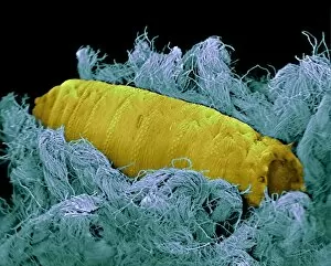
Blowfly pupa on carpet. Coloured scanning electron micrograph (SEM) of a Calliphora vicina blowfly pupa that will hatch an adult fly. A blowfly lays its eggs on dead bodies

Background imageFibres Collection: Ant sensory hairs, SEM

Ant sensory hairs, SEM
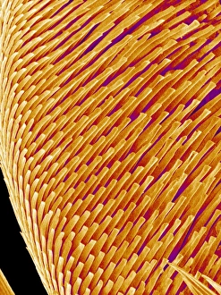
Ant sensory hairs. Coloured scanning electron micrograph (SEM) of an ants sensory hairs. These are bristles that are growing through the ants exoskeleton

Background imageFibres Collection: Sperm tails, TEM

Sperm tails, TEM
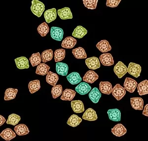
Sperm tails. Coloured transmission electron micrograph (TEM) of cross sections through sperm tails. A sperms tails ability to move is partly due to the arrangement of microtubules

Background imageFibres Collection: Periodontal ligament fibres, SEM

Periodontal ligament fibres, SEM
Periodontal ligament fibres. Coloured scanning electron micrograph (SEM) of periodontal ligament fibres (red). These fibres, a specialised type of connective tissue

Background imageFibres Collection: Collagen fibres, TEM

Collagen fibres, TEM
Collagen fibres. Coloured transmission electron micrograph (TEM) of collagen protein fibres. Collagen has a high tensile strength, providing structure and elasticity to skin, tendons

Background imageFibres Collection: Connective tissue fibres

Connective tissue fibres
Connective tissue. False-colour scanning electron micrograph (SEM) of bundles of collagen and elastic fibres which form part of the connective tissue

Background imageFibres Collection: Neural connective tissue, SEM

Neural connective tissue, SEM
Neural connective tissue. Scanning electron micrograph (SEM) of collagen bundles forming the delicate connective tissue called endoneurium

Background imageFibres Collection: Coloured SEM of collagen connective tissue fibres

Coloured SEM of collagen connective tissue fibres
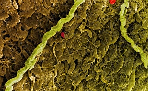
Collagen connective tissue. Coloured scanning electron micrograph (SEM) of connective tissue formed by bundles of wavy collagen fibres. Red blood cells (red) are also seen

Background imageFibres Collection: Collagen bundles, SEM

Collagen bundles, SEM
Collagen bundles. Scanning electron micrograph (SEM) of collagen bundles from the delicate connective tissue endoneurium. Endoneurium wraps around and between individual nerve fibres (axons)

Background imageFibres Collection: Artwork of collagen fibres in connective tissue

Artwork of collagen fibres in connective tissue
Artwork representing collagen fibres. They are found throughout the connective tissue which provides structural and metabolic support for other tissues and organs throughout the body

Background imageFibres Collection: Ear wax, SEM

Ear wax, SEM
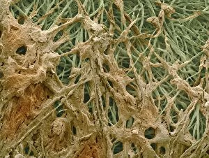
Ear wax. Coloured scanning electron micrograph (SEM) of ear wax (brown) collected on cotton bud fibres (green). Ear wax forms in the ear canal, which runs between the outer ear and the ear drum

Background imageFibres Collection: Myelin surrounding a nerve axon, TEM

Myelin surrounding a nerve axon, TEM
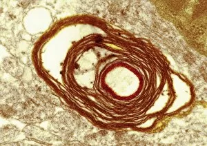
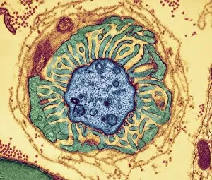
Myelin surrounding a nerve axon, coloured transmission electron micrograph (TEM). The concentric round rings are the sheets of a Schwann cells myelin membrane (brown rings)

Background imageFibres Collection: Retina, SEM

Retina, SEM
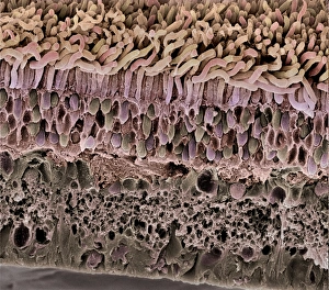
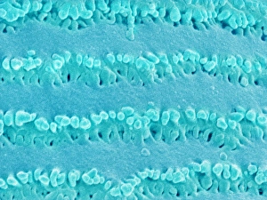
Retina. Coloured scanning electron micrograph (SEM) of a section through a freeze-fractured retina, showing the light-sensitive rods and cones and their associated neurones

Background imageFibres Collection: Lens fibre of an eye

Lens fibre of an eye
Lens fibre from an eye. Coloured scanning electron micrograph (SEM) of the fibre-like cells that form the crystalline lens of the eye

Background imageFibres Collection: Cornea, SEM

Cornea, SEM
Cornea. Coloured scanning electron micrograph (SEM) of a section through a cornea, the transparent region on the outer surface of the eye. The front of the eye is at top

Background imageFibres Collection: Skeletal muscle fibre

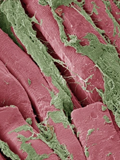
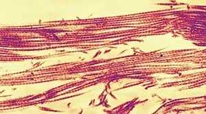
Skeletal muscle fibre. Coloured scanning electron micrograph (SEM) of skeletal muscle fibre. This type of muscle is striated

Background imageFibres Collection: Smooth muscle, SEM

Smooth muscle, SEM
Smooth muscle. Coloured scanning electron micrograph of smooth muscle from the trachea (windpipe). Smooth muscle is not under voluntary control, unlike skeletal (striated) muscle

Background imageFibres Collection: Contracted artery, TEM

Contracted artery, TEM
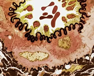
Contracted elastic artery. Coloured transmission electron micrograph (TEM) of a section through an artery. Red blood cells (erythrocytes) are seen in the lumen (top centre)

Background imageFibres Collection: Somatic peripheral nervous system

Somatic peripheral nervous system
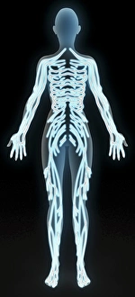
Peripheral nervous system. Computer artwork of the main nerves of the somatic part of the peripheral nervous system. The peripheral nervous system does not include the brain and spinal cord

Background imageFibres Collection: Surgical face mask, SEM

Surgical face mask, SEM
Surgical face mask, coloured scanning electron micrograph (SEM). This mask is made of the plant material cellulose, which is biodegradable

Background imageFibres Collection: Straw particleboard

Straw particleboard. Piece of particleboard made from wheat straw. Particles of straw are produced by milling bales of waste straw, and then bound by a resin to produce a wood-like building material

Background imageFibres Collection: Banknote security

Banknote security. Euro banknote in ultraviolet light. This highlights security features intended to prevent fraud. Fluorescent ink and strips glow bright blue and red under ultraviolet light

Background imageFibres Collection: False-colour SEM of cells from lens of hu

False-colour SEM of cells from lens of hu

Background imageFibres Collection: Myelinated nerve fibres, SEM

Myelinated nerve fibres, SEM
Myelinated nerve fibres. Coloured scanning electron micrograph (SEM) of a section through a bundle of nerve fibres containing a blood vessel (orange)

Background imageFibres Collection: Neurovascular bundle, TEM

Neurovascular bundle, TEM
Neurovascular bundle. Coloured transmission electron micrograph (TEM) of a cross section through a neurovascular bundle (NVB)

Background imageFibres Collection: Nerve fibre node, TEM

Nerve fibre node, TEM
Nerve fibre node. Coloured transmission electron micrograph (TEM) of a cross-section through a nerve fibre (axon) at a node of Ranvier

Background imageFibres Collection: Nerve fibres, TEM

Nerve fibres, TEM
Nerve fibres. Coloured transmission electron micrograph (TEM) of a section through non- myelinated nerve fibres (blue). The fibres are arranged into bundles surrounded by the cytoplasm of Schwann


All Professionally Made to Order for Quick Shipping

"Fibres: Unraveling the Intricacies of the Human Brain and Beyond" Exploring the intricate network of brain fibres through DTI MRI scans (C017 / 7099). The mesmerizing artwork depicting white matter fibres and their connection to the brain (C015 / 1930). Delving deeper into understanding brain fibres with DTI MRI scans (C017 / 7035). Illuminating neurons and astrocytes through immunofluorescent LM, revealing the complexity of brain fibres. Peering into the white matter fibres of the human brain, unlocking its mysteries (C014 / 5666). Witnessing nerve cells regenerate under a TEM microscope, showcasing nature's resilience. Marveling at the intricate structure of a lime tree stem captured in a light micrograph. Discovering beauty in unexpected places - castor oil stem captured in a mesmerizing light micrograph. Unveiling myelination's role in enhancing nerve fibre function through TEM imaging. Witnessing cell division unfold under fluorescent micrographs, highlighting life's continuous renewal process. Celebrating nature's ability to heal as we observe regenerating nerve cells under TEM microscopy once again. Exploring further into white matter fibres within our complex human brains (C014/5668). In this captivating journey, we unravel the wonders – from delicate neural connections within our brains to resilient plant stems that provide sustenance for life itself.