Fraud Blocker Skip to main content

Fibres Collection (page 3)

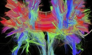
"Fibres: Unraveling the Intricacies of the Human Brain and Beyond" Exploring the intricate network of brain fibres through DTI MRI scans (C017 / 7099

Background imageFibres Collection: Scutching or dressing flax by beating the stalks by hand. The long fibres of the stem Flax plant
Scutching or dressing flax by beating the stalks by hand. The long fibres of the stem Flax plant (Linum) were processed to produce linen

Background imageFibres Collection: Rippling flax to remove the seeds and remains of flower heads. In the background

Rippling flax to remove the seeds and remains of flower heads. In the background the flax plants are being pulled (harvested)

Background imageFibres Collection: The Great Hall of Marshalls Flax Mill, Leeds, Yorkshire, England. The roof is

The Great Hall of Marshalls Flax Mill, Leeds, Yorkshire, England. The roof is supported on cast iron columns, and glazed skylights give even overhead light

Background imageFibres Collection: Loaf of rye bread being wrapped in brown paper, side paper

Loaf of rye bread being wrapped in brown paper, side paper

Background imageFibres Collection: Fresh green cabbage

Fresh green cabbage

Background imageFibres Collection: Three carrots with full green shoots and three others with cropped shoots, front view

Three carrots with full green shoots and three others with cropped shoots, front view

Background imageFibres Collection: Selection of vegetable leaves, including Pak Choi, Rocket, Chicory, Frisee and Radicchio

Selection of vegetable leaves, including Pak Choi, Rocket, Chicory, Frisee and Radicchio

Background imageFibres Collection: Two heads of green cabbage, front view

Two heads of green cabbage, front view

Background imageFibres Collection: Wholewheat cereal biscuit, close up, view from above

Wholewheat cereal biscuit, close up, view from above

Background imageFibres Collection: Bowl of oat cereal and fresh strawberries, blackberries and raspberries with milk

Bowl of oat cereal and fresh strawberries, blackberries and raspberries with milk, laid on top of plate with napkin and spoon, glass and jug of water in background, front view

Background imageFibres Collection: Selection of vegetable leaves, including Savoy Cabbage, Pak Choi, Red Chard, Chinese Leaf and Rocket

Selection of vegetable leaves, including Savoy Cabbage, Pak Choi, Red Chard, Chinese Leaf and Rocket

Background imageFibres Collection: Bowl of cornflakes, view from above

Bowl of cornflakes, view from above

Background imageFibres Collection: Selection of foods, half a pomegranate, red pear, cabbage head

Selection of foods, half a pomegranate, red pear, cabbage head, partially sliced loaf of wholemeal bread, whole fish, carrots, turnip, pulses and grains

Background imageFibres Collection: Fresh Celery leafstalks

Fresh Celery leafstalks

Background imageFibres Collection: Glass of muesli topped with yoghurt, peach slices and raspberries, front view

Glass of muesli topped with yoghurt, peach slices and raspberries, front view

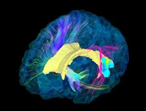
Background imageFibres Collection: Corpus callosum, DTI MRI scan C017 / 7044

Corpus callosum, DTI MRI scan C017 / 7044
Corpus callosum. Coronal 3D diffusion tensor imaging (DTI) magnetic resonance imaging (MRI) scan of nerve pathways (coloured) in and around the brains corpus callosum

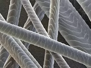
Background imageFibres Collection: Domestic cat hair, coloured SEM Z934 / 0486

Domestic cat hair, coloured SEM Z934 / 0486
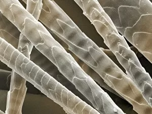
Cat hair. Coloured scanning electron micrograph (SEM) of the hair from a domestic cat (Felis domesticus). Magnification: x370 when printed 10cm high

Background imageFibres Collection: Grey squirrel hair, coloured SEM

Grey squirrel hair, coloured SEM
Grey squirrel hair. Coloured scanning electron micrograph (SEM) of the hair from a Grey squirrel (Sciurus carolinensis). Magnification: x730 when printed 10cm wide

Background imageFibres Collection: Wild rabbit hair, coloured SEM

Wild rabbit hair, coloured SEM
Wild rabbit hair. Coloured scanning electron micrograph (SEM) of the hair from a wild rabbit (Oryctolagus cuniculus). Magnification: x360 when printed 10cm high

Background imageFibres Collection: Camel hair, coloured SEM Z949 / 0010

Camel hair, coloured SEM Z949 / 0010
Camel hair. Coloured scanning electron micrograph (SEM) of the hair from a dromedary camel. These short dense hairs allow the camel to survive the extreme temperatures of the desert by reflecting

Background imageFibres Collection: Rabbit hair, coloured SEM Z938 / 0037

Rabbit hair, coloured SEM Z938 / 0037
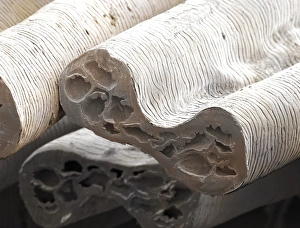
Rabbit hair. Coloured scanning electron micrograph (SEM) of the transverse section of hair from a rabbit (Oryctolagus cuniculus). Magnification: x670 when printed 10cm high

Background imageFibres Collection: Horse body hair, coloured SEM

Horse body hair, coloured SEM
Horse hair. Coloured scanning electron micrograph (SEM) of hair from the body of a horse (Equus caballus). Magnification: x300 when printed 10cm high

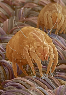
Background imageFibres Collection: Dust mites

Dust mites. Coloured scanning electron micrograph (SEM) of two dust mites (Dermatophagoides sp.) on fabric fibres. Millions of dust mites inhabit the home, feeding on shed skin cells

Background imageFibres Collection: Red squirrel hair, coloured SEM

Red squirrel hair, coloured SEM
Red squirrel hair. Coloured scanning electron micrograph (SEM) of the hair from a red squirrel (Sciurus vulgaris). Magnification: x370 when printed 10cm high

Background imageFibres Collection: Horse mane hair, coloured SEM

Horse mane hair, coloured SEM
Horse hair. Coloured scanning electron micrograph (SEM) of hair from the mane of a horse (Equus caballus). Magnification: x2000 when printed 10cm high

Background imageFibres Collection: Mouse hair, coloured SEM Z918 / 0198

Mouse hair, coloured SEM Z918 / 0198
House mouse hair. Coloured scanning electron micrograph (SEM) of hair from a house mouse (Mus musculus). Magnification: x900 when printed 10cm wide

Background imageFibres Collection: Grey seal hair, coloured SEM

Grey seal hair, coloured SEM
Grey seal hair. Coloured scanning electron micrograph of the hair from a grey seal (Halichoerus grypus). Magnification: x161 when printed at 10cm height

Background imageFibres Collection: Hedgehog hair, coloured SEM

Hedgehog hair, coloured SEM
Hedgehog hair. Coloured scanning electron micrograph of the hairs from a hedgehog (Erinaceus europaeus). Magnification: x130 when printed 10cm high

Background imageFibres Collection: Horse tail hair, coloured SEM

Horse tail hair, coloured SEM
Horse hair. Coloured scanning electron micrograph (SEM) of hair from the tail of a horse (Equus caballus). Magnification: x200 when printed 10cm high

Background imageFibres Collection: Torn postage stamp, SEM

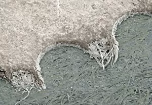
Torn postage stamp, SEM
Torn postage stamp. Coloured scanning electron micrograph (SEM) of a torn perforation from the edge of a postage stamp. Magnification: x26 at 6x7cm size

Background imageFibres Collection: Household dust, SEM

Household dust, SEM
Household dust, coloured scanning electron micrograph (SEM). Magnification: x130 when printed at 10 centimetres wide

Background imageFibres Collection: Electric blanket heating element, SEM

Electric blanket heating element, SEM
Heating element. Coloured scanning electron micrograph (SEM) of part of the heating element from an electric blanket. A wire (gold)

Background imageFibres Collection: Silkworm silk fibres (SEM) C014 / 2150

Silkworm silk fibres (SEM) C014 / 2150
Silkworm silk fibres, coloured scanning electron micrograph (SEM). The silkworm is the larva or caterpillar of the silkmoth which has been domesticated from the wild silkmoth Bombyx mandarina

Background imageFibres Collection: Brain fibres, DTI MRI scan C017 / 7037

Brain fibres, DTI MRI scan C017 / 7037
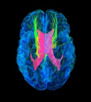
Brain fibres. 3D diffusion tensor imaging (DTI) magnetic resonance imaging (MRI) scan of a selection of nerve pathways (green/yellow) in the brain

Background imageFibres Collection: Brain fibres, DTI MRI scan C017 / 7038

Brain fibres, DTI MRI scan C017 / 7038
Brain fibres. 3D diffusion tensor imaging (DTI) magnetic resonance imaging (MRI) scan of a selection of nerve pathways (green/yellow) in the brain

Background imageFibres Collection: Brain tumour, DTI MRI scan C017 / 7057

Brain tumour, DTI MRI scan C017 / 7057
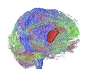
Brain tumour. 3D diffusion tensor imaging (DTI) magnetic resonance imaging (MRI) scan of nerve pathways (coloured) in a brain with a tumour (red, centre right)

Background imageFibres Collection: Brain fibres, DTI MRI scan C017 / 7100

Brain fibres, DTI MRI scan C017 / 7100
Brain fibres. 3D diffusion tensor imaging (DTI) magnetic resonance imaging (MRI) scan of a selection of nerve pathways (blue, pink, green) in the brain. The front of the brain is at left

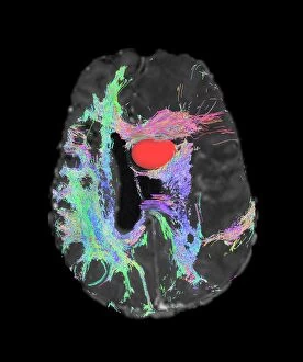
Background imageFibres Collection: Brain tumour, DTI MRI scan C017 / 7059

Brain tumour, DTI MRI scan C017 / 7059
Brain tumour. Axial diffusion tensor imaging (DTI) magnetic resonance imaging (MRI) scan of nerve pathways (coloured) in a brain with a tumour (red, upper centre)

Background imageFibres Collection: Brain tumour, DTI MRI scan C017 / 7058

Brain tumour, DTI MRI scan C017 / 7058
Brain tumour. 3D diffusion tensor imaging (DTI) magnetic resonance imaging (MRI) scan of nerve pathways (coloured) in a brain with a tumour (red, centre left)

Background imageFibres Collection: Sugar uptake in muscles, diagram

Sugar uptake in muscles, diagram
Sugar uptake in muscles. Diagram showing sugar from a drink (upper right) being taken up by the muscles of the human body

Background imageFibres Collection: Peripheral nerve, light micrograph

Peripheral nerve, light micrograph
Peripheral nerve. Light micrograph of a section through a peripheral nerve. This is a mixed nerve with myelinated axons (dark blue circles)

Background imageFibres Collection: Nerve ganglion, light micrograph

Nerve ganglion, light micrograph
Nerve ganglion. Light micrograph of a section through a dorsal (sensory) spinal root ganglion associated with a sensory nerve root of the spinal cord. Sensory information from peripheral sites e.g

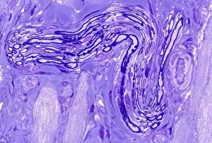
Background imageFibres Collection: Myelinated nerve, light micrograph

Myelinated nerve, light micrograph
Myelinated nerve. Light micrograph of a section through a peripheral myelinated nerve, showing many individual axons each covered with deep blue-staining myelin sheaths

Background imageFibres Collection: Structure of the cochlea, artwork

Structure of the cochlea, artwork
Structure of the cochlea. Computer artwork of a section through the organ of Corti, the auditory sense organ that lines the spiral of the cochlea in the inner ear

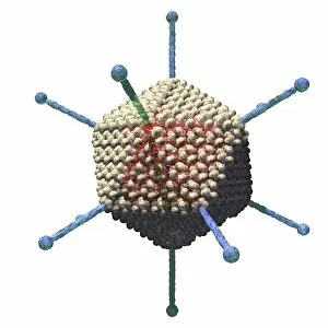
Background imageFibres Collection: Adenovirus, artwork C016 / 8960

Adenovirus, artwork C016 / 8960
Adenovirus. Computer artwork showing the external protein structure of an adenovirus particle. Adenoviruses are the largest viruses not to have a protein coat covering their capsid

Background imageFibres Collection: Adenovirus, artwork C016 / 8961

Adenovirus, artwork C016 / 8961
Adenovirus. Computer artwork showing the external protein structure of an adenovirus particle. Adenoviruses are the largest viruses not to have a protein coat covering their capsid

Background imageFibres Collection: Adenovirus particles, artwork C016 / 8963

Adenovirus particles, artwork C016 / 8963
Adenovirus particles. Computer artwork showing the external protein structure of adenovirus particles. Adenoviruses are the largest viruses not to have a protein coat covering their capsid

Background imageFibres Collection: Adenovirus particles, artwork C016 / 8965

Adenovirus particles, artwork C016 / 8965
Adenovirus particles. Computer artwork showing the external protein structure of adenovirus particles. Adenoviruses are the largest viruses not to have a protein coat covering their capsid


All Professionally Made to Order for Quick Shipping

"Fibres: Unraveling the Intricacies of the Human Brain and Beyond" Exploring the intricate network of brain fibres through DTI MRI scans (C017 / 7099). The mesmerizing artwork depicting white matter fibres and their connection to the brain (C015 / 1930). Delving deeper into understanding brain fibres with DTI MRI scans (C017 / 7035). Illuminating neurons and astrocytes through immunofluorescent LM, revealing the complexity of brain fibres. Peering into the white matter fibres of the human brain, unlocking its mysteries (C014 / 5666). Witnessing nerve cells regenerate under a TEM microscope, showcasing nature's resilience. Marveling at the intricate structure of a lime tree stem captured in a light micrograph. Discovering beauty in unexpected places - castor oil stem captured in a mesmerizing light micrograph. Unveiling myelination's role in enhancing nerve fibre function through TEM imaging. Witnessing cell division unfold under fluorescent micrographs, highlighting life's continuous renewal process. Celebrating nature's ability to heal as we observe regenerating nerve cells under TEM microscopy once again. Exploring further into white matter fibres within our complex human brains (C014/5668). In this captivating journey, we unravel the wonders – from delicate neural connections within our brains to resilient plant stems that provide sustenance for life itself.