Fraud Blocker Skip to main content

Fibres Collection (page 9)

"Fibres: Unraveling the Intricacies of the Human Brain and Beyond" Exploring the intricate network of brain fibres through DTI MRI scans (C017 / 7099

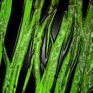
Background imageFibres Collection: Muscle fibres

Muscle fibres. Computer-enhanced confocal light micrograph of skeletal (striated) muscle fibres. Skeletal muscle is under the conscious control of the brain

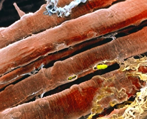
Background imageFibres Collection: Coloured SEM of skeletal (striated) muscle fibres

Coloured SEM of skeletal (striated) muscle fibres
Skeletal muscle. Coloured scanning electron micrograph (SEM) of skeletal muscle fibres. Muscle fibres are extremely long, cylindrical cells

Background imageFibres Collection: Coloured SEM of a bundle of skeletal muscle fibres

Coloured SEM of a bundle of skeletal muscle fibres

Background imageFibres Collection: Brain

Brain, historical artwork. The top drawing shows the brain cut in-between its hemispheres. The front of the brain is on the left

Background imageFibres Collection: Alzheimers disease brain cell, TEM

Alzheimers disease brain cell, TEM
Alzheimers disease brain cell. Coloured transmission electron micrograph (TEM) of a neurofibrillary tangle in a nerve cell from the brain of a patient with Alzheimers disease

Background imageFibres Collection: Moving lights, abstract image

Moving lights, abstract image

Background imageFibres Collection: Fibres in a bra strap, SEM

Fibres in a bra strap, SEM
Fibres in a bra strap. Coloured scanning electron micrograph (SEM) of the fibres in a bra. These are mainly nylon and polyester

Background imageFibres Collection: Fibres in a bra, SEM

Fibres in a bra, SEM
Fibres in a bra. Coloured scanning electron micrograph (SEM) of the fibres in a bra. These are mainly nylon and polyester

Background imageFibres Collection: Woven wool, SEM

Woven wool, SEM
Woven wool. Coloured scanning electron micrograph (SEM) of fibres of wool woven together

Background imageFibres Collection: Polypropylene filaments, SEM

Polypropylene filaments, SEM
Polypropylene filaments. Coloured scanning electron micrograph (SEM) of filaments of the plastic polypropylene, formed by the polymerization of propylene

Background imageFibres Collection: Man made coloured fibres

Man made coloured fibres

Background imageFibres Collection: Woven fabric, SEM

Woven fabric, SEM
Woven fabric, coloured scanning electron micrograph (SEM)

Background imageFibres Collection: Dirty scouring pad

Dirty scouring pad. Coloured scanning electron micrograph (SEM) of a dirty scouring pad. Particles of food (green) trapped in the pads fibres (yellow)

Background imageFibres Collection: Collecting evidence

Collecting evidence. Forensic officer using a suction tube device to collect evidence from the seat of a car. This tool is used to collect fibres and hairs for later laboratory analysis

Background imageFibres Collection: Wool, SEM

Wool, SEM
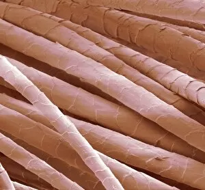
Wool. Coloured scanning electron micrograph (SEM) of wool from a sheep. The cuticle (outer layer) of the hair comprises overlapping scales

Background imageFibres Collection: Net curtain, light micrograph

Net curtain, light micrograph
Net curtain, polarised light micrograph. This curtain is made from synthetic fibres

Background imageFibres Collection: Cotton and synthetic fibres, SEM

Cotton and synthetic fibres, SEM
Cotton and synthetic underwear fibres. Coloured scanning electron micrograph (SEM) of a cotton and polyurethane mixture of fibres woven to form part of a womans underwear item

Background imageFibres Collection: Synthetic fibres, SEM

Synthetic fibres, SEM
Synthetic fibres. Coloured scanning electron micrograph (SEM) of synthetic fibres woven to form material for clothing

Background imageFibres Collection: Woven fabric fibres, SEM

Woven fabric fibres, SEM
Woven fabric fibres. Coloured scanning electron micrograph (SEM) of fibres woven into a lattice of interlocking parts. This is part of a cloth called Georgette crepe

Background imageFibres Collection: Phase change material, SEM

Phase change material, SEM
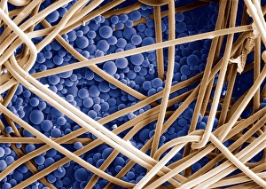
Phase change material. Coloured scanning electron micrograph (SEM) of microcapsules (blue) that contain a phase change material (PCM) coating fabric fibres

Background imageFibres Collection: SEM of non-woven Nylon geotextile fibres

SEM of non-woven Nylon geotextile fibres
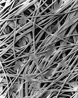
Nylon geotextile. Scanning electron micrograph of fibres of nylon geotextile. This material consists of non-woven fibres which are thermally bonded

Background imageFibres Collection: Eye lens, SEM

Eye lens, SEM
Eye lens. Coloured scanning electron micrograph (SEM) of cells from the lens (crystalline lens) of the eye. The cells are in a stacked linear, fibre-like arrangement

Background imageFibres Collection: Seaweed paper fibres, SEM

Seaweed paper fibres, SEM
Seaweed paper fibres, coloured scanning electron micrograph (SEM). Many plant fibres, including seaweed (marine algae), can be used to make paper. Magnification: x30 at 6x7cm size

Background imageFibres Collection: Filter paper, SEM

Filter paper, SEM
Filter paper. Scanning electron micrograph (SEM) of the cut end of a piece of filter paper showing its many cellulose fibres

Background imageFibres Collection: Stressed culture cell, SEM

Stressed culture cell, SEM

Background imageFibres Collection: Magazine paper, SEM

Magazine paper, SEM
Magazine paper, coloured scanning electron micrograph (SEM). Paper mostly consists of a mesh of softened and pulped wood fibres

Background imageFibres Collection: Bone structure, macrophotograph

Bone structure, macrophotograph
Bone structure. Macrophotograph of a model of normal human bone structure

Background imageFibres Collection: Used surgical swab, SEM

Used surgical swab, SEM
Used surgical swab, coloured scanning electron micrograph (SEM). Red blood cells (erythrocytes, red) and fibrin (purple) are stuck to the fibres of the swab

Background imageFibres Collection: Toothbrush bristles, SEM

Toothbrush bristles, SEM
Toothbrush bristles, coloured scanning electron micrograph (SEM). These bristles are designed to be used in tooth brushing to remove food debris and dental plaque from teeth

Background imageFibres Collection: Nerve fibres, light micrograph

Nerve fibres, light micrograph
Nerve fibres. Light micrograph of a transverse section through myelinated nerve fibres. The myelin sheaths are black, axons are blue. Magnification: x100 when printed at 10 centimetres wide

Background imageFibres Collection: Skeletal muscle fibres, SEM

Skeletal muscle fibres, SEM
Skeletal muscle fibres, coloured scanning electron micrograph (SEM). Endomysial connective tissue is green. Magnification: x300 when printed at 10 centimetres wide

Background imageFibres Collection: Nerve fibres, SEM

Nerve fibres, SEM
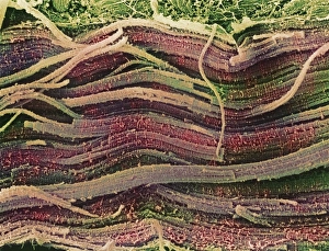
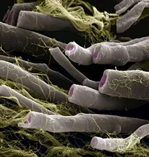
Myelinated nerve fibres, coloured scanning electron micrograph (SEM). The myelin sheath is grey, the axoplasm pink and the endoneurium (connective tissue) yellow

Background imageFibres Collection: Cashmere wool, SEM

Cashmere wool, SEM
Cashmere wool. Coloured scanning electron micrograph (SEM) of hair fibres from the cashmere goat. The cuticle (outer layer) of the hair comprises overlapping scales

Background imageFibres Collection: Clothes moth, SEM

Clothes moth, SEM
Clothes moth. Coloured scanning electron micrograph (SEM) of a common clothes moth (Tineola bisselliella) on cashmere wool fibres

Background imageFibres Collection: Adenovirus structure, artwork

Adenovirus structure, artwork
Adenovirus structure. Computer artwork showing the external protein structure of a human adenovirus (HadV) particle. Adenoviruses are the largest viruses not to have a protein coat covering their

Background imageFibres Collection: Synthetic fibre, light micrograph

Synthetic fibre, light micrograph
Light micrograph of a synthetic fibre, taken with polarised light. Magnification: x100

Background imageFibres Collection: Spinal cord, SEM

Spinal cord, SEM
Spinal cord. Coloured scanning electron micrograph (SEM) of a section through a spinal cord, showing the grey matter (green) and white matter (blue)

Background imageFibres Collection: Optic nerve fibres, SEM

Optic nerve fibres, SEM
Optic nerve fibres. Coloured scanning electron micrograph (SEM) of axons from the optic nerve. Connective tissue fibres (thread-like) are also visible

Background imageFibres Collection: Ageing skin, artwork

Ageing skin, artwork
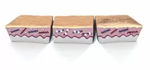
Ageing skin. Computer artwork of sections through human skin, showing its appearance as it ages (from left to right). Elastin (red) and collagen (blue) in the skins dermis (pink) break down

Background imageFibres Collection: Cells and fibres, historical artwork

Cells and fibres, historical artwork. Early 20th Century illustrations of various cells and fibres. Published in Quains dictionary of medicine (3rd edition), by the Irish physician

Background imageFibres Collection: Coconut rope production, 19th century

Coconut rope production, 19th century
Coconut rope production, 19th-century artwork. The fibres for this rope are obtained from the husk of the coconut, producing a material known as coir

Background imageFibres Collection: Carbon fibres, SEM

Carbon fibres, SEM
Carbon fibres, coloured scanning electron micrograph (SEM). These fibres can be woven into a fabric that is strong, but lightweight

Background imageFibres Collection: African teak woody stem, light micrograph

African teak woody stem, light micrograph
African teak woody stem. Polarised light micrograph of a section through the stem of an African teak (Pericopsis elata) plant, showing its diffuse-porous xylem tissue

Background imageFibres Collection: Fat tissue, SEM

Fat tissue, SEM
Fat tissue. Coloured scanning electron micrograph (SEM) of fat cells (adipocytes, round) surrounded by fine strands of supportive connective tissue


All Professionally Made to Order for Quick Shipping

"Fibres: Unraveling the Intricacies of the Human Brain and Beyond" Exploring the intricate network of brain fibres through DTI MRI scans (C017 / 7099). The mesmerizing artwork depicting white matter fibres and their connection to the brain (C015 / 1930). Delving deeper into understanding brain fibres with DTI MRI scans (C017 / 7035). Illuminating neurons and astrocytes through immunofluorescent LM, revealing the complexity of brain fibres. Peering into the white matter fibres of the human brain, unlocking its mysteries (C014 / 5666). Witnessing nerve cells regenerate under a TEM microscope, showcasing nature's resilience. Marveling at the intricate structure of a lime tree stem captured in a light micrograph. Discovering beauty in unexpected places - castor oil stem captured in a mesmerizing light micrograph. Unveiling myelination's role in enhancing nerve fibre function through TEM imaging. Witnessing cell division unfold under fluorescent micrographs, highlighting life's continuous renewal process. Celebrating nature's ability to heal as we observe regenerating nerve cells under TEM microscopy once again. Exploring further into white matter fibres within our complex human brains (C014/5668). In this captivating journey, we unravel the wonders – from delicate neural connections within our brains to resilient plant stems that provide sustenance for life itself.