Fraud Blocker Skip to main content

Imaging Technique Collection

"Unveiling the Intricacies of Brain Fibres

Background imageImaging Technique Collection: Brain fibres, DTI MRI scan C017 / 7099

Brain fibres, DTI MRI scan C017 / 7099
Brain fibres. 3D diffusion tensor imaging (DTI) magnetic resonance imaging (MRI) scan of nerve pathways in the brain. The pathways are highlighted in green and blue. The brain is seen from the front

Background imageImaging Technique Collection: Brain fibres, DTI MRI scan C017 / 7035

Brain fibres, DTI MRI scan C017 / 7035
Brain fibres. 3D diffusion tensor imaging (DTI) magnetic resonance imaging (MRI) scan of nerve pathways in the brain. The pathways are highlighted in blue

Background imageImaging Technique Collection: Shuttle Test Using Electron Beam

Shuttle Test Using Electron Beam
A space shuttle model undergoes a wind tunnel test in 1975. This test is simulating the ionized gasses that surround a shuttle as it reenters the atmosphere

Background imageImaging Technique Collection: Brain tumour, fMRI and tractography C017 / 7102

Brain tumour, fMRI and tractography C017 / 7102
Brain tumour, fMRI and tractography. Combined functional magnetic resonance imaging (fMRI, blue and green) and tractography (yellow and red) imaging of a brain with a tumour (upper left)

Background imageImaging Technique Collection: Corticospinal tract, DTI MRI scan C017 / 7046

Corticospinal tract, DTI MRI scan C017 / 7046
Corticospinal tract. Sagittal 3D diffusion tensor imaging (DTI) magnetic resonance imaging (MRI) scan of nerve pathways (blue) in the brains corticospinal tract

Background imageImaging Technique Collection: Brain fibres, tract density imaging C017 / 7039

Brain fibres, tract density imaging C017 / 7039
Brain fibres, tract density imaging. Axial scan of the brain showing the density of nerve pathways (tracts) in the brain. The image was obtained using 3D diffusion tensor imaging (DTI)

Background imageImaging Technique Collection: Corpus callosum, DTI MRI scan C017 / 7043

Corpus callosum, DTI MRI scan C017 / 7043
Corpus callosum. Coronal 3D diffusion tensor imaging (DTI) magnetic resonance imaging (MRI) scan of nerve pathways (red) in the brains corpus callosum

Background imageImaging Technique Collection: Brains white matter, DTI MRI scan

Brains white matter, DTI MRI scan
Brains white matter. Close-up of an area of the brain imaged using tract density imaging and 3D diffusion tensor imaging (DTI), a magnetic resonance imaging (MRI) technique

Background imageImaging Technique Collection: Brain tumour, DTI modelling C017 / 7060

Brain tumour, DTI modelling C017 / 7060
Brain tumour. Computer artwork of nerve pathways (coloured) in a brain with a tumour (white/red, upper right). The brain is seen from the front. Brain tumours can be benign or malignant (cancerous)

Background imageImaging Technique Collection: Brain fibres, DTI MRI scan C017 / 7036

Brain fibres, DTI MRI scan C017 / 7036
Brain fibres. 3D diffusion tensor imaging (DTI) magnetic resonance imaging (MRI) scan of a selection of nerve pathways (green/yellow) in the brain

Background imageImaging Technique Collection: Artery damage, MRA scan

Artery damage, MRA scan
Artery damage. Magnetic resonance angiography (MRA) scan of the neck of a 23-year-old male motorcycle crash victim. Dissection of the right internal carotid artery (left)

Background imageImaging Technique Collection: Corpus callosum, DTI MRI scan C017 / 7044

Corpus callosum, DTI MRI scan C017 / 7044
Corpus callosum. Coronal 3D diffusion tensor imaging (DTI) magnetic resonance imaging (MRI) scan of nerve pathways (coloured) in and around the brains corpus callosum

Background imageImaging Technique Collection: Brain fibres, DTI MRI scan C017 / 7037

Brain fibres, DTI MRI scan C017 / 7037
Brain fibres. 3D diffusion tensor imaging (DTI) magnetic resonance imaging (MRI) scan of a selection of nerve pathways (green/yellow) in the brain

Background imageImaging Technique Collection: Brain fibres, DTI MRI scan C017 / 7038

Brain fibres, DTI MRI scan C017 / 7038
Brain fibres. 3D diffusion tensor imaging (DTI) magnetic resonance imaging (MRI) scan of a selection of nerve pathways (green/yellow) in the brain

Background imageImaging Technique Collection: Brain tumour, DTI MRI scan C017 / 7057

Brain tumour, DTI MRI scan C017 / 7057
Brain tumour. 3D diffusion tensor imaging (DTI) magnetic resonance imaging (MRI) scan of nerve pathways (coloured) in a brain with a tumour (red, centre right)

Background imageImaging Technique Collection: Brain fibres, DTI MRI scan C017 / 7100

Brain fibres, DTI MRI scan C017 / 7100
Brain fibres. 3D diffusion tensor imaging (DTI) magnetic resonance imaging (MRI) scan of a selection of nerve pathways (blue, pink, green) in the brain. The front of the brain is at left

Background imageImaging Technique Collection: Brain tumour, DTI MRI scan C017 / 7059

Brain tumour, DTI MRI scan C017 / 7059
Brain tumour. Axial diffusion tensor imaging (DTI) magnetic resonance imaging (MRI) scan of nerve pathways (coloured) in a brain with a tumour (red, upper centre)

Background imageImaging Technique Collection: Brain tumour, DTI MRI scan C017 / 7058

Brain tumour, DTI MRI scan C017 / 7058
Brain tumour. 3D diffusion tensor imaging (DTI) magnetic resonance imaging (MRI) scan of nerve pathways (coloured) in a brain with a tumour (red, centre left)

Background imageImaging Technique Collection: Corpus callosum, DTI MRI scan C017 / 7045

Corpus callosum, DTI MRI scan C017 / 7045
Corpus callosum. Coronal 3D diffusion tensor imaging (DTI) magnetic resonance imaging (MRI) scan of nerve pathways (blue) in and around the brains corpus callosum

Background imageImaging Technique Collection: Glioblastoma brain tumour, DTI MRI scan C017 / 7052

Glioblastoma brain tumour, DTI MRI scan C017 / 7052
Glioblastoma brain tumour. 3D diffusion tensor imaging (DTI) magnetic resonance imaging (MRI) scan of nerve pathways (coloured) around a glioblastoma (upper right) in the brain

Background imageImaging Technique Collection: Brain injury, MRI scan

Brain injury, MRI scan
Brain injury. Coronal magnetic resonance imaging (MRI) brain scan showing intracranial abnormalities in a 23-year-old male motorcycle crash victim

Background imageImaging Technique Collection: Fluorescence lifetime imaging lasers

Fluorescence lifetime imaging lasers. Lasers of a fluorescence lifetime imaging (FLIM) microscope in a medical research laboratory


All Professionally Made to Order for Quick Shipping

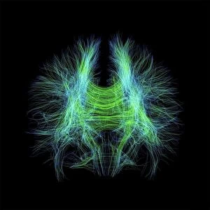
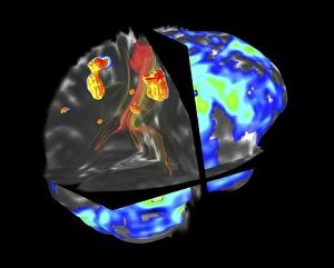
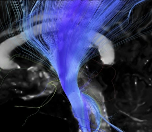
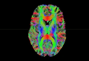
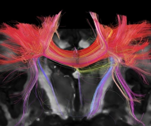
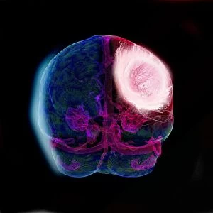
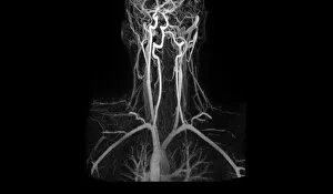
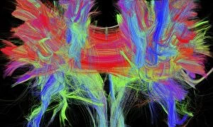
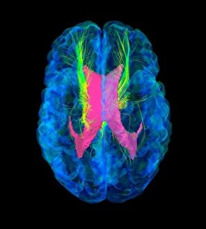
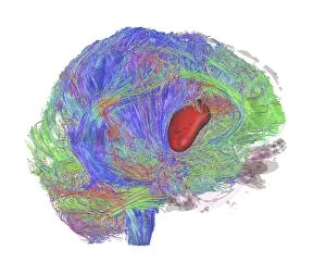
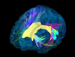
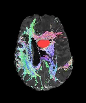
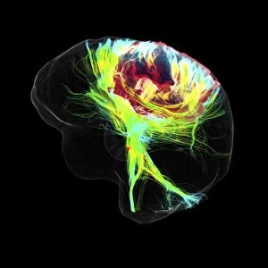
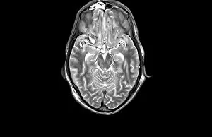
"Unveiling the Intricacies of Brain Fibres: Exploring Imaging Techniques" Discovering the complex network of brain fibres has become increasingly possible with advanced imaging techniques. The DTI MRI scan C017 / 7099 and C017 / 7035 have allowed researchers to delve into the intricate pathways within our brains, shedding light on their connectivity. Innovative methods such as the Shuttle Test Using Electron Beam have provided a unique perspective on brain function, enabling scientists to observe how different regions communicate and interact. When it comes to diagnosing brain tumours, fMRI and tractography have proven invaluable. The C017 / 7102 scan combines these techniques, offering detailed insights into tumour growth patterns and potential treatment options. The Corticospinal tract is another area of interest that has been extensively studied using DTI MRI scans (C017 / 7046). By visualizing this crucial pathway responsible for motor control, researchers gain a better understanding of conditions affecting movement. Tract density imaging (C017 / 7039) further enhances our knowledge of brain fibres by providing information about their distribution throughout white matter. This technique allows us to map out connections in unprecedented detail. Artery damage can also be assessed through MRA scans, helping identify potential risks or causes behind certain neurological conditions. Glioblastoma brain tumours pose significant challenges in diagnosis and treatment planning. However, DTI MRI scans (C017 / 7048 & C017/7055) offer valuable data regarding tumor location and its impact on surrounding structures. As we continue exploring imaging techniques like never before, we unlock new possibilities for understanding the complexities of our brains. These advancements hold immense promise for improving diagnoses accuracy and developing targeted treatments tailored to each individual's unique neural architecture.