
Neural Tract Collection
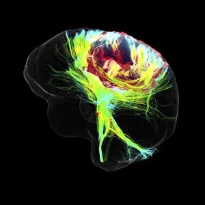
"Unraveling the Intricacies of Neural Tracts: Insights from Advanced MRI Scans" The human brain is a complex network of interconnected fibers


All Professionally Made to Order for Quick Shipping
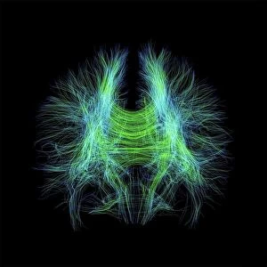
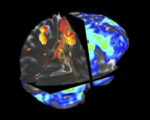
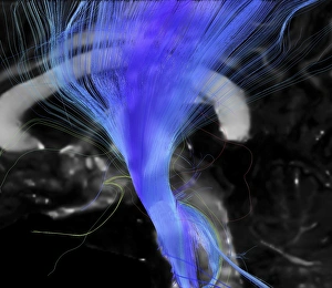
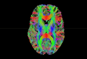
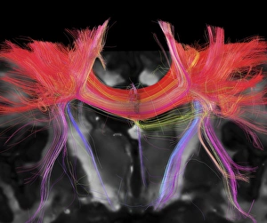
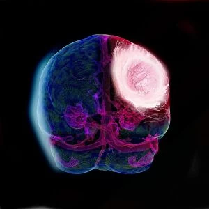
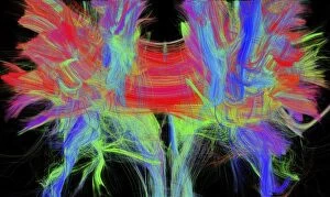
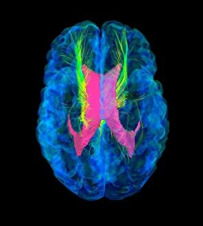
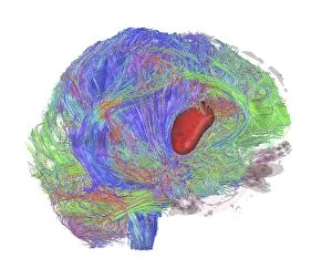
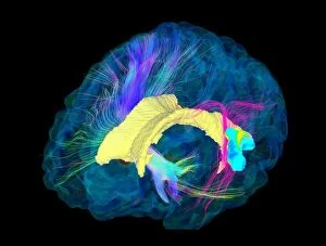
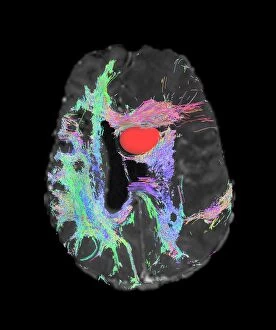
"Unraveling the Intricacies of Neural Tracts: Insights from Advanced MRI Scans" The human brain is a complex network of interconnected fibers, responsible for transmitting vital information throughout our bodies. Thanks to cutting-edge technology like DTI (Diffusion Tensor Imaging) MRI scans, we can now delve deeper into understanding these neural tracts. In image C017 / 7099 and C017 / 7035, we witness the intricate web of brain fibers illuminated by DTI MRI scans. This non-invasive technique allows us to visualize the structural integrity and connectivity within the brain's white matter. Moving beyond mere visualization, fMRI (functional Magnetic Resonance Imaging) combined with tractography provides valuable insights into conditions such as brain tumors. In scan C017 / 7102, we observe how this powerful combination helps map out neural pathways affected by a tumor. This knowledge aids in surgical planning and treatment decisions. Specifically focusing on the corticospinal tract, scan C017 / 7046 showcases how DTI MRI enables precise mapping of this crucial pathway involved in motor function. Such detailed imaging assists medical professionals in diagnosing and monitoring neurological disorders more accurately. Tract density imaging further enhances our understanding of brain fiber organization, as demonstrated in image C017 / 7039. By quantifying fiber density along specific tracts, researchers gain valuable insights into various cognitive processes and neurological conditions. Glioblastoma brain tumors pose significant challenges due to their aggressive nature. However, through advanced techniques like DTI modeling showcased in images C017 / 7060, C017 / 7048, C017 /7055 &C107/7056), we can better comprehend tumor growth patterns and their impact on surrounding neural structures. One critical structure that benefits from DTI analysis is the corpus callosum – connecting both hemispheres of the brain.