Fraud Blocker Skip to main content

Brain Scan Collection

"Unraveling the Intricacies of Brain Pathways

Background imageBrain Scan Collection: Brain pathways

Brain pathways

Background imageBrain Scan Collection: Brain fibres, DTI MRI scan C017 / 7099

Brain fibres, DTI MRI scan C017 / 7099
Brain fibres. 3D diffusion tensor imaging (DTI) magnetic resonance imaging (MRI) scan of nerve pathways in the brain. The pathways are highlighted in green and blue. The brain is seen from the front

Background imageBrain Scan Collection: Brain fibres, DTI MRI scan C017 / 7035

Brain fibres, DTI MRI scan C017 / 7035
Brain fibres. 3D diffusion tensor imaging (DTI) magnetic resonance imaging (MRI) scan of nerve pathways in the brain. The pathways are highlighted in blue

Background imageBrain Scan Collection: White matter fibres of the human brain C014 / 5666

White matter fibres of the human brain C014 / 5666
White matter fibres. Coloured 3D diffusion spectral imaging (DSI) scan of the bundles of white matter nerve fibres in the brain

Background imageBrain Scan Collection: White matter fibres of the human brain C014 / 5668

White matter fibres of the human brain C014 / 5668
White matter fibres. Coloured 3D diffusion spectral imaging (DSI) scan of the bundles of white matter nerve fibres in the brain

Background imageBrain Scan Collection: White matter fibres of the human brain C014 / 5667

White matter fibres of the human brain C014 / 5667
White matter fibres. Coloured 3D diffusion spectral imaging (DSI) scan of the bundles of white matter nerve fibres in the brain

Background imageBrain Scan Collection: Healthy brain, MRI scans

Healthy brain, MRI scans
Healthy brain. Coloured magnetic resonance imaging (MRI) scans through the head of a 55 year old man, showing the brain sectioned in coronal (frontal) view

Background imageBrain Scan Collection: Childs brain, MRI scan

Childs brain, MRI scan
Childs brain. Coloured magnetic resonance imaging (MRI) scan of an coronal (vertical) section through the head and brain of a healthy 3-year-old child, passing through the ears

Background imageBrain Scan Collection: White matter fibres, DTI scan

White matter fibres, DTI scan
White matter fibres. Coloured 3D diffusion tensor imaging (DTI) scan of the brain, showing the bundles of white matter nerve fibres

Background imageBrain Scan Collection: Brain tumour, fMRI and tractography C017 / 7102

Brain tumour, fMRI and tractography C017 / 7102
Brain tumour, fMRI and tractography. Combined functional magnetic resonance imaging (fMRI, blue and green) and tractography (yellow and red) imaging of a brain with a tumour (upper left)

Background imageBrain Scan Collection: Corticospinal tract, DTI MRI scan C017 / 7046

Corticospinal tract, DTI MRI scan C017 / 7046
Corticospinal tract. Sagittal 3D diffusion tensor imaging (DTI) magnetic resonance imaging (MRI) scan of nerve pathways (blue) in the brains corticospinal tract

Background imageBrain Scan Collection: Brain fibres, tract density imaging C017 / 7039

Brain fibres, tract density imaging C017 / 7039
Brain fibres, tract density imaging. Axial scan of the brain showing the density of nerve pathways (tracts) in the brain. The image was obtained using 3D diffusion tensor imaging (DTI)

Background imageBrain Scan Collection: Corpus callosum, DTI MRI scan C017 / 7043

Corpus callosum, DTI MRI scan C017 / 7043
Corpus callosum. Coronal 3D diffusion tensor imaging (DTI) magnetic resonance imaging (MRI) scan of nerve pathways (red) in the brains corpus callosum

Background imageBrain Scan Collection: Brains white matter, DTI MRI scan

Brains white matter, DTI MRI scan
Brains white matter. Close-up of an area of the brain imaged using tract density imaging and 3D diffusion tensor imaging (DTI), a magnetic resonance imaging (MRI) technique

Background imageBrain Scan Collection: Brain tumour, DTI modelling C017 / 7060

Brain tumour, DTI modelling C017 / 7060
Brain tumour. Computer artwork of nerve pathways (coloured) in a brain with a tumour (white/red, upper right). The brain is seen from the front. Brain tumours can be benign or malignant (cancerous)

Background imageBrain Scan Collection: MRI brain scan F006 / 9208

MRI brain scan F006 / 9208
Brain scan. False-colour magnetic resonance imaging (MRI) scan of a human head containing a healthy brain, seen in horizontal view. At upper frame, the two eyeballs can be seen

Background imageBrain Scan Collection: Brain fibres, DTI MRI scan C017 / 7036

Brain fibres, DTI MRI scan C017 / 7036
Brain fibres. 3D diffusion tensor imaging (DTI) magnetic resonance imaging (MRI) scan of a selection of nerve pathways (green/yellow) in the brain

Background imageBrain Scan Collection: White matter fibres of the human brain C014 / 5670

White matter fibres of the human brain C014 / 5670
White matter fibres. Coloured 3D diffusion spectral imaging (DSI) scan of the bundles of white matter nerve fibres in the brain

Background imageBrain Scan Collection: Brain MRI scan with Alzheimers QR code

Brain MRI scan with Alzheimers QR code
White matter fibres overlaid a mri scan of the human head. Coloured 3D diffusion spectral imaging (DSI) scan of the bundles of white matter nerve fibres in the brain

Background imageBrain Scan Collection: White matter fibres, brain mri scan C014 / 5674

White matter fibres, brain mri scan C014 / 5674
White matter fibres overlaid a mri scan of the human head. Coloured 3D diffusion spectral imaging (DSI) scan of the bundles of white matter nerve fibres in the brain

Background imageBrain Scan Collection: Brain tumour, 3D-MRI scan

Brain tumour, 3D-MRI scan
Brain tumour. Coloured three-dimensional model of a male head, made up from numerous magnetic resonance imaging (MRI) scans

Background imageBrain Scan Collection: Telemedicine, conceptual image C017 / 7605

Telemedicine, conceptual image C017 / 7605
MODEL RELEASED. Telemedicine, conceptual image

Background imageBrain Scan Collection: Telemedicine, conceptual image C017 / 7603

Telemedicine, conceptual image C017 / 7603
MODEL RELEASED. Telemedicine, conceptual image

Background imageBrain Scan Collection: Brain scan, conceptual image C017 / 7392

Brain scan, conceptual image C017 / 7392
MODEL RELEASED. Brain scan, conceptual composite image

Background imageBrain Scan Collection: Brain scan, conceptual image C017 / 7394

Brain scan, conceptual image C017 / 7394
MODEL RELEASED. Brain scan, conceptual composite image

Background imageBrain Scan Collection: Brain scan, conceptual image C017 / 7398

Brain scan, conceptual image C017 / 7398
MODEL RELEASED. Brain scan, conceptual composite image

Background imageBrain Scan Collection: Brain scan, conceptual image C017 / 7397

Brain scan, conceptual image C017 / 7397
MODEL RELEASED. Brain scan, conceptual composite image

Background imageBrain Scan Collection: Corpus callosum, DTI MRI scan C017 / 7044

Corpus callosum, DTI MRI scan C017 / 7044
Corpus callosum. Coronal 3D diffusion tensor imaging (DTI) magnetic resonance imaging (MRI) scan of nerve pathways (coloured) in and around the brains corpus callosum

Background imageBrain Scan Collection: Brain scan, conceptual image C017 / 7391

Brain scan, conceptual image C017 / 7391
MODEL RELEASED. Brain scan, conceptual composite image

Background imageBrain Scan Collection: Coloured MRI scan of the human head F007 / 4202

Coloured MRI scan of the human head F007 / 4202
Brain scan. False-colour magnetic resonance imaging (MRI) scan of a human head containing a healthy brain, seen in horizontal view. At upper frame, the two eyeballs can be seen

Background imageBrain Scan Collection: Brain scan, MRI scan, heightmap F006 / 7096

Brain scan, MRI scan, heightmap F006 / 7096
Normal brain. Coloured magnetic resonance imaging (MRI) scan of an axial section through a healthy brain, converted into a heightmap or height field

Background imageBrain Scan Collection: Brain scan, MRI scan, heightmap F006 / 7080

Brain scan, MRI scan, heightmap F006 / 7080
Normal brain. Coloured magnetic resonance imaging (MRI) scan of an axial section through a healthy brain, converted into a heightmap or height field

Background imageBrain Scan Collection: Brain scan, MRI scan F006 / 4629

Brain scan, MRI scan F006 / 4629
Normal brain. Coloured magnetic resonance imaging (MRI) scan of an axial section through a healthy brain. The front of the brain is towards top of the image

Background imageBrain Scan Collection: Brain scan, MRI scan F006 / 4610

Brain scan, MRI scan F006 / 4610
Normal brain. Coloured magnetic resonance imaging (MRI) scan of an axial section through a healthy brain. The front of the brain is towards top of the image

Background imageBrain Scan Collection: Brain scan, MRI scan F006 / 4617

Brain scan, MRI scan F006 / 4617
Normal brain. Coloured magnetic resonance imaging (MRI) scan of an axial section through a healthy brain. The front of the brain is towards top of the image

Background imageBrain Scan Collection: MRI brain scan and nerve cells F006 / 3595

MRI brain scan and nerve cells F006 / 3595
Computer artwork of a sagittal (side) view of a human head. The largest part of the brain is the cerebrum (upper part). This surrounds the limbic system (curved structures at upper centre)

Background imageBrain Scan Collection: Brain scan, MRI scan F006 / 4607

Brain scan, MRI scan F006 / 4607
Normal brain. Coloured magnetic resonance imaging (MRI) scan of an axial section through a healthy brain. The front of the brain is towards top of the image

Background imageBrain Scan Collection: Brain and nerve cells, neural network F006 / 3585

Brain and nerve cells, neural network F006 / 3585
Computer artwork of a frontal view of a human brain. In the background a neural network of nerve cells firing

Background imageBrain Scan Collection: Brain scan, MRI scan, heightmap F006 / 7078

Brain scan, MRI scan, heightmap F006 / 7078
Normal brain. Coloured magnetic resonance imaging (MRI) scan of an axial section through a healthy brain, converted into a heightmap or height field

Background imageBrain Scan Collection: Brain scan, MRI scan, heightmap F006 / 7093

Brain scan, MRI scan, heightmap F006 / 7093
Normal brain. Coloured magnetic resonance imaging (MRI) scan of an axial section through a healthy brain, converted into a heightmap or height field

Background imageBrain Scan Collection: Brain scan, MRI scan, heightmap F006 / 7081

Brain scan, MRI scan, heightmap F006 / 7081
Normal brain. Coloured magnetic resonance imaging (MRI) scan of an axial section through a healthy brain, converted into a heightmap or height field

Background imageBrain Scan Collection: Brain scan, MRI scan, heightmap F006 / 7091

Brain scan, MRI scan, heightmap F006 / 7091
Normal brain. Coloured magnetic resonance imaging (MRI) scan of an axial section through a healthy brain, converted into a heightmap or height field

Background imageBrain Scan Collection: Brain haemorrhage, MRI scan F008 / 3466

Brain haemorrhage, MRI scan F008 / 3466
Brain haemorrhage. Magnetic resonance imaging (MRI) scan of the brain of a 74 year old female WHO has had a massive intracranial haemorrhage (yellow)

Background imageBrain Scan Collection: Brain haemorrhage, MRI scan F008 / 3468

Brain haemorrhage, MRI scan F008 / 3468
Brain haemorrhage. Magnetic resonance imaging (MRI) scan of the brain of a 74 year old female who has had a massive intracranial haemorrhage (red)

Background imageBrain Scan Collection: Thought process, artwork F005 / 0195

Thought process, artwork F005 / 0195
Thought process, conceptual computer artwork, showing a brain gear in a human head profile

Background imageBrain Scan Collection: Thought process, artwork F005 / 0192

Thought process, artwork F005 / 0192
Thought process, conceptual computer artwork, showing a brain gear overlaid a brain model

Background imageBrain Scan Collection: Coloured MRI scan of the human head F007 / 4205

Coloured MRI scan of the human head F007 / 4205
Brain scan. False-colour magnetic resonance imaging (MRI) scan of a human head containing a healthy brain, seen in horizontal view. At upper frame, the two eyeballs can be seen

Background imageBrain Scan Collection: Brain fibres, DTI MRI scan C017 / 7037

Brain fibres, DTI MRI scan C017 / 7037
Brain fibres. 3D diffusion tensor imaging (DTI) magnetic resonance imaging (MRI) scan of a selection of nerve pathways (green/yellow) in the brain


All Professionally Made to Order for Quick Shipping

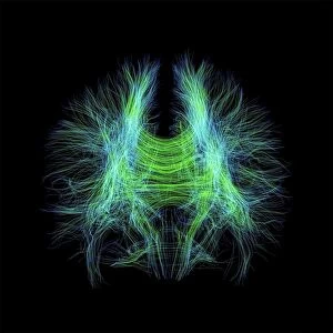
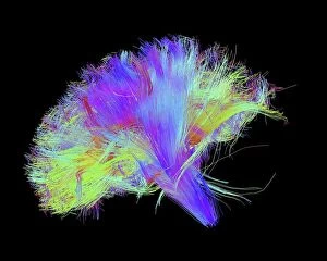
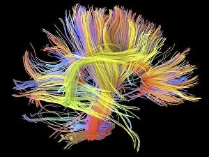
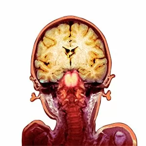
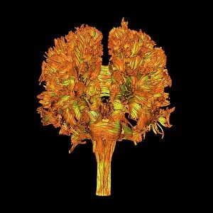
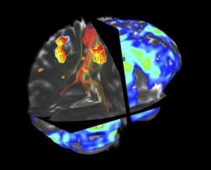
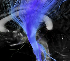
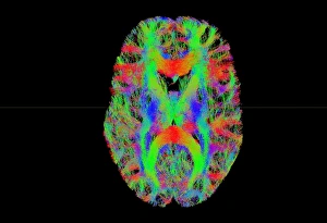
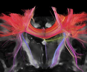
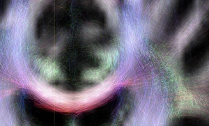
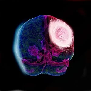
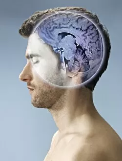
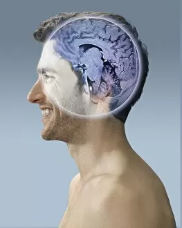
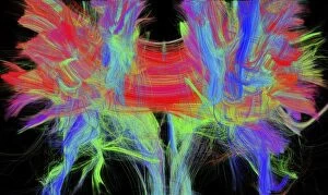
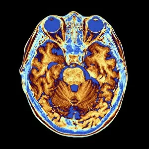
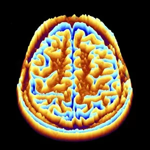
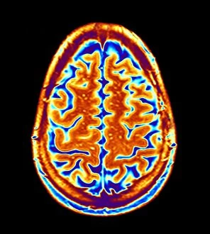
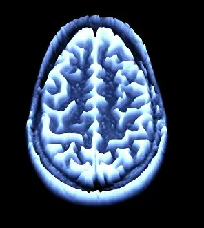
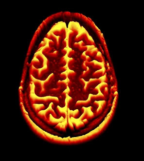
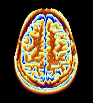
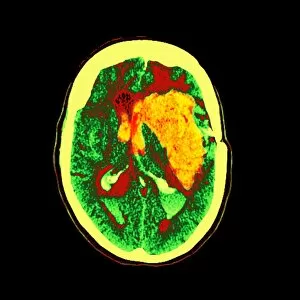
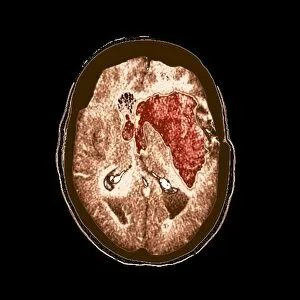
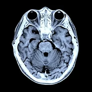
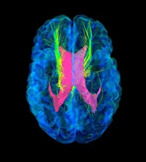
"Unraveling the Intricacies of Brain Pathways: A Glimpse into the Fascinating World of Brain Scans" Discovering the hidden secrets within our minds has always been a captivating endeavor. With advancements in technology, we now have an unprecedented opportunity to delve deeper into the intricate network of brain fibres and pathways. Through cutting-edge techniques like DTI MRI scans (C017 / 7099), scientists are unraveling the mysteries that lie beneath. The white matter fibres of the human brain (C014 / 5666) hold invaluable clues about how information is transmitted throughout this remarkable organ. By examining these fibres using DTI MRI scans (C017 / 7035), researchers can map out complex connections and gain insights into cognitive processes. Intriguingly, these investigations extend beyond just healthy brains; they also shed light on neurological disorders such as Alzheimer's disease. Comparisons between normal and Alzheimer's-affected brains offer glimpses into how this devastating condition disrupts vital brain functions. Moreover, even young minds are not exempt from scrutiny. Childs' brain MRI scans provide valuable data for understanding early development and identifying potential abnormalities or delays that may require intervention. One particularly striking example lies in coloured MRI scans showcasing organophosphate brain damage. These images serve as a stark reminder of the consequences certain substances can have on our delicate neural networks, urging us to prioritize safeguarding our mental well-being. As we continue to explore further horizons within neuroscience, each new scan brings us closer to unlocking profound discoveries about ourselves and what it means to be human. The realm of brain imaging holds immense promise for revolutionizing healthcare by enabling earlier diagnoses, personalized treatments, and ultimately enhancing our understanding of consciousness itself. So let us marvel at these mesmerizing snapshots – snapshots that capture both beauty and complexity – as they guide us towards a future where we harness the power of knowledge to unlock the full potential of our most extraordinary organ: the human brain.