
Structural Collection
"Unveiling the Intricate Web: Exploring Structural Wonders from Brain Fibres to DNA Molecules" Delving into the Complexity


All Professionally Made to Order for Quick Shipping
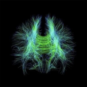
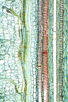
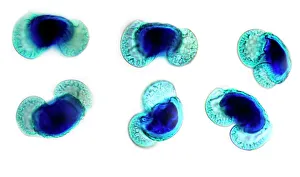
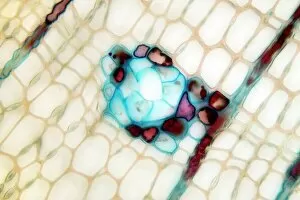
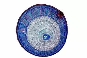
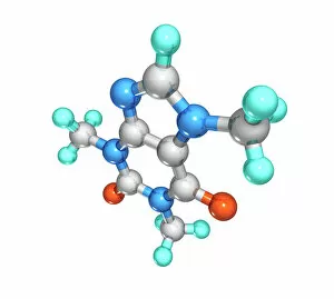
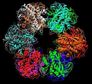
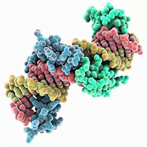
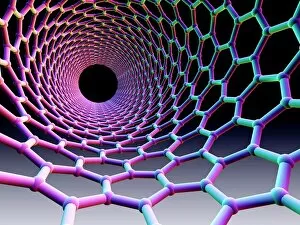
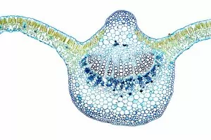
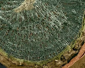
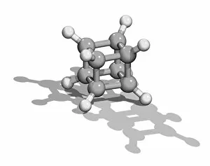
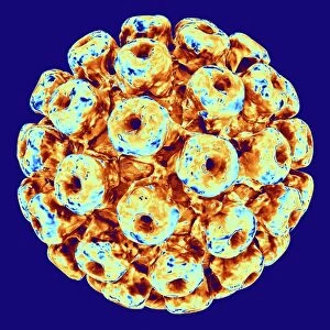
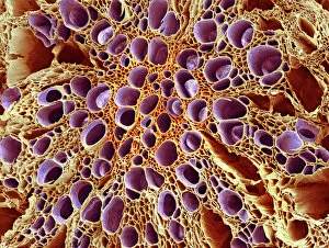
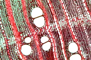
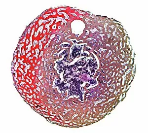
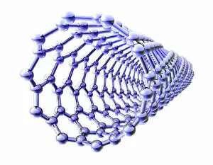
"Unveiling the Intricate Web: Exploring Structural Wonders from Brain Fibres to DNA Molecules" Delving into the Complexity: DTI MRI scan C017 / 7099 reveals the intricate network of brain fibres, offering a glimpse into our cognitive architecture. Unraveling Mysteries: DTI MRI scan C017 / 7035 uncovers hidden connections within brain fibres, shedding light on how information flows through our neural pathways. Soaring Through History: The Hawker Hurricane Fighter, 1939 stands as a testament to the structural brilliance that revolutionized aerial warfare. Nature's Blueprint Revealed: A captivating light micrograph showcases the delicate structure of a dicotyledon plant stem, highlighting its role in supporting growth and nourishment. X-ray Artistry at its Finest: Skeletons come alive in mesmerizing artwork created through X-rays, capturing both their structural beauty and enigmatic allure. Building Blocks of Life: Dive deep into cellular wonders with an awe-inspiring glimpse into cell structures that form the foundation of all living organisms. Beneath the Surface Beauty: Witness skeletal marvels from below through stunning X-ray artwork that unveils hidden intricacies and celebrates human anatomy. Unlocking Genetic Secrets: A computer model presents a captivating visualization of DNA molecules, showcasing their structural elegance and vital role in heredity. Pollen's Dance in Light: Pine pollen grains take center stage under a microscope's gaze, revealing their exquisite architectural design crafted by nature itself. Illuminating Botanical Marvels: Explore the enchanting world within lime tree stems through a mesmerizing light micrograph that captures their unique structural patterns and resilience. Caffeine's Molecular Symphony : Journey inside caffeine's molecular realm as we uncover its intricate structure – fuel for millions around the globe.