
Tractogram Collection
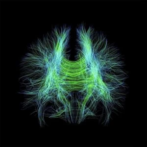
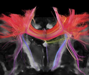
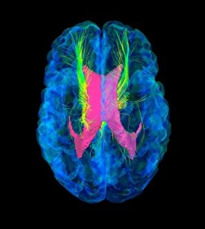
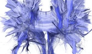
Exploring the intricate pathways of brain fibres through DTI MRI scans C017 / 7099 and C017 / 7035, revealing the complexity within our minds


All Professionally Made to Order for Quick Shipping
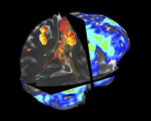
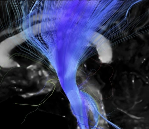
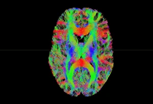
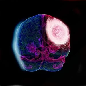
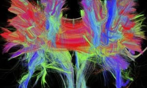
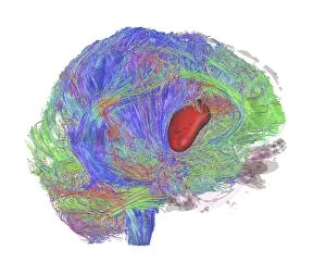
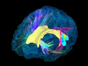
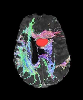
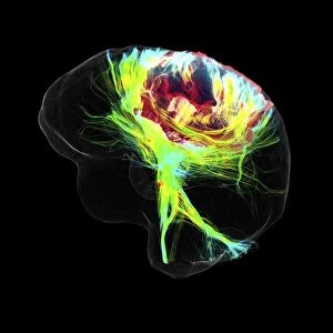
Exploring the intricate pathways of brain fibres through DTI MRI scans C017 / 7099 and C017 / 7035, revealing the complexity within our minds. Unveiling the secrets hidden by a brain tumour using fMRI and tractography in scan C017 / 7102. Delving into the Corticospinal tract's structure with precision through DTI MRI scan C017 / 7046, shedding light on its vital role in motor function. Tract density imaging uncovers the vast network of brain fibres in scan C017 / 7039, showcasing the interconnectedness of our thoughts and actions. Witnessing the wonders of white matter through DTI MRI scans, unraveling new insights about cognitive processes. Examining glioblastoma brain tumours' impact on neural connections via DTI modelling in scan C017 / 7060, providing crucial information for treatment strategies. Further exploration into brain fibres with DTI MRI scans C017 / 7036 reveals more about their organization and functionality. Gaining deeper understanding into glioblastoma brain tumours' effects on cognition through DTI MRI scans C017/7048, C017/7055, and C107/7056; paving way for improved therapies. The Corpus callosum's significance comes to light as we analyze its integrity using DTI MRI scan C107/7044; highlighting its role in interhemispheric communication.