
Mri Scanner Collection
"Exploring the Intricacies of Brain Fibres: A Journey through MRI Scans" Delving deep into the complexities of brain fibres


All Professionally Made to Order for Quick Shipping
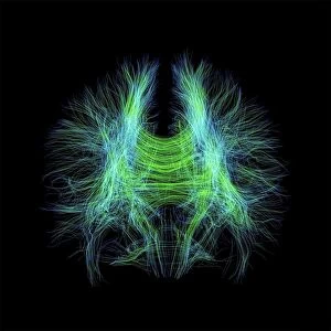
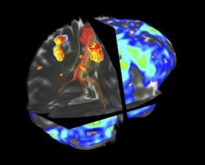
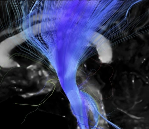
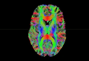
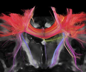
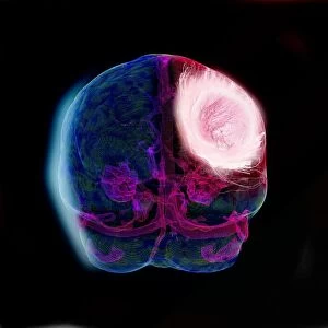
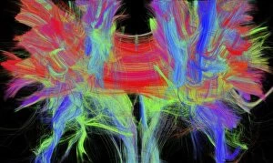
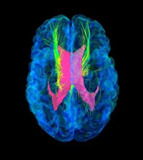
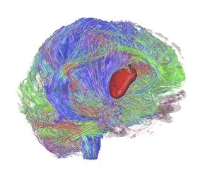
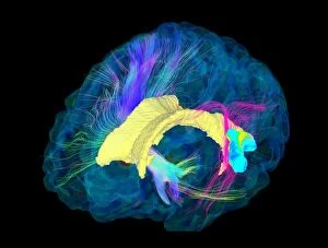
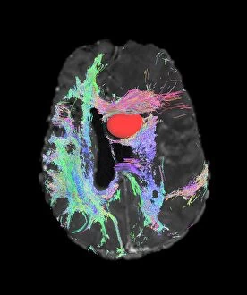
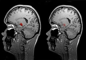
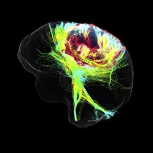
"Exploring the Intricacies of Brain Fibres: A Journey through MRI Scans" Delving deep into the complexities of brain fibres, DTI MRI scans C017 / 7099 and C017 / 7035 unveil a mesmerizing world within our minds. These advanced imaging techniques provide us with invaluable insights into brain anatomy, allowing us to comprehend its intricate structure. In the realm of medical diagnostics, fMRI and tractography C017 / 7102 prove indispensable in detecting brain tumours. By combining functional magnetic resonance imaging (fMRI) with tractography, doctors can accurately visualize and map out the affected areas for precise treatment planning. The corticospinal tract takes center stage in DTI MRI scan C017 / 7046, shedding light on this vital pathway responsible for motor control. This revelation enhances our understanding of how signals travel from the brain to different parts of the body. Tract density imaging presents another breakthrough as showcased in C017 / 7039, enabling researchers to study brain fibres' distribution across various regions. Such knowledge contributes significantly to unraveling mysteries surrounding white matter connectivity within our brains. DTI MRI scans offer unparalleled glimpses into both healthy and diseased brains alike. Through DTI modelling captured in C017 / 7060, we gain valuable insights into how tumours affect neural pathways—empowering medical professionals with crucial information for effective treatment strategies. As we explore further using DTI MRI scan C017 / 7036, an awe-inspiring cross-sectional biomedical illustration reveals a remarkable plane that dissects through layers of grey matter—a testament to human ingenuity merging with technological advancements. Glioblastoma brain tumour studies take precedence as seen in DTI MRI scans C017 / 7048 and C017/7055. These images expose the devastating impact these tumors have on delicate neural networks while inspiring researchers worldwide to find innovative solutions against this formidable adversary.