
Cerebrum Collection
The cerebrum, the largest part of the human brain, is a fascinating structure that plays a crucial role in our cognitive abilities


All Professionally Made to Order for Quick Shipping
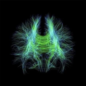
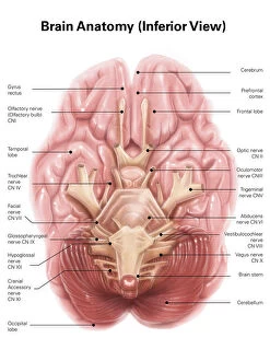
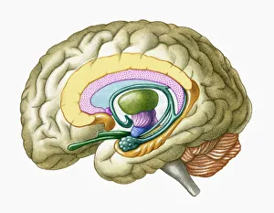
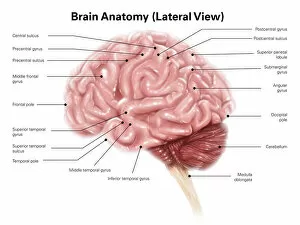
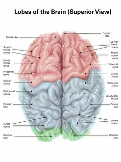
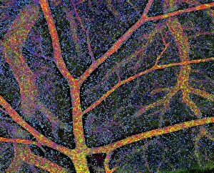
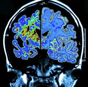
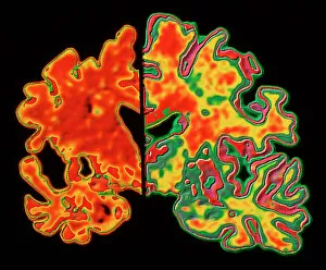
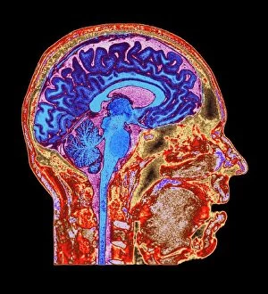
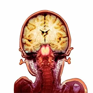
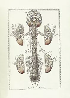
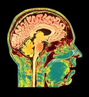
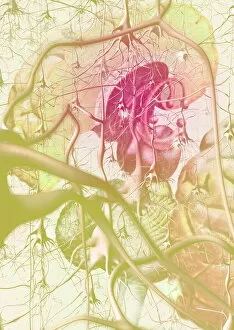
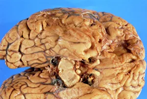
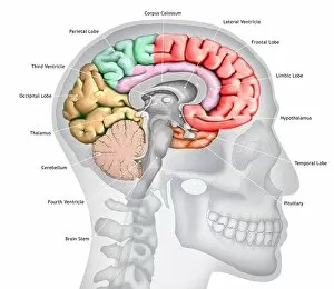
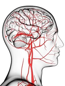
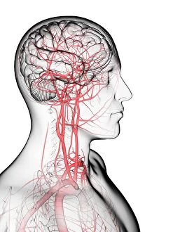
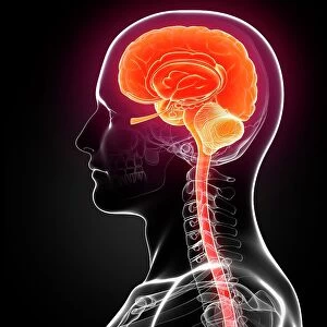
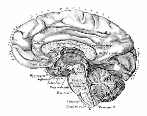
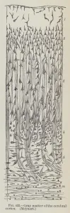
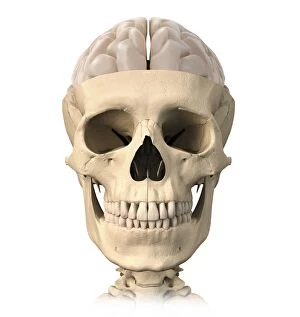
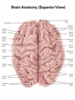
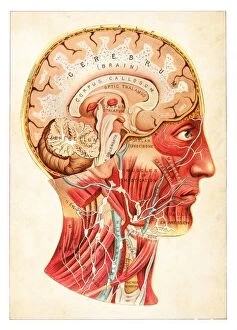
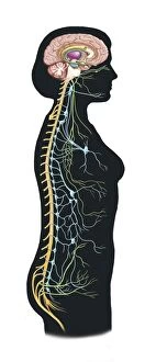
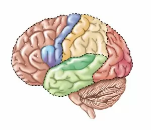
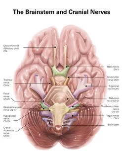
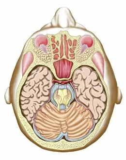
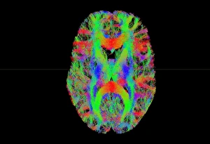
The cerebrum, the largest part of the human brain, is a fascinating structure that plays a crucial role in our cognitive abilities. In this captivating image, we get an inferior view of the anatomy of the cerebrum, showcasing its intricate network of brain fibres captured through a DTI MRI scan (C017 / 7099). These brain pathways are like highways for information transmission within our brains. Another DTI MRI scan (C017 / 7035) provides us with a closer look at these remarkable brain fibres. It's truly astonishing how these delicate structures enable communication between different regions of the cerebrum. A cross-section illustration reveals not only the limbic system but also gives us insight into the primitive forebrain. This primitive region has evolved over time and contributes to our emotions and instincts. From a superior view, we can observe colored lobes labeled on this human brain. Each lobe serves distinct functions such as motor control, sensory perception, language processing, and more. The complexity and organization within this organ are awe-inspiring. Moving to a lateral view of human brain anatomy highlights its various structures and their interconnectedness. This perspective allows us to appreciate how each component works together seamlessly to facilitate our everyday activities. An MRI scan showcases both normalcy (C016 / 8845) and Alzheimer's-affected brains. Through computer artwork depicting Alzheimer's disease progression in the brain tissue, we gain insight into how this devastating condition impacts neural connections over time. Lastly, understanding blood supply to cerebral tissues is vital for comprehending overall brain function. By studying it closely alongside other images related to Alzheimer's disease or general anatomy scans helps researchers uncover potential links between vascular health and neurological disorders. Exploring these captivating images sheds light on different aspects anatomy - from its complex fiber networks captured through advanced imaging techniques like DTI MRI scans to illustrations highlighting specific regions like limbic system and primitive forebrain.