
Neural Pathway Collection
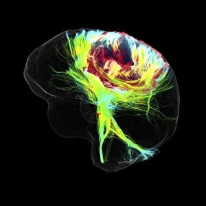
"Unraveling the Intricacies of Neural Pathways: A Journey into the Brain's Fibres" The human brain, with its complex network of neural pathways


All Professionally Made to Order for Quick Shipping
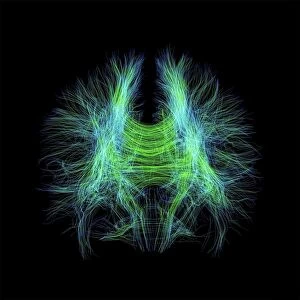
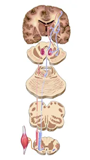
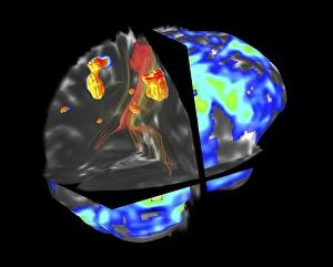
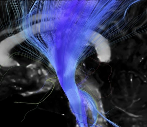
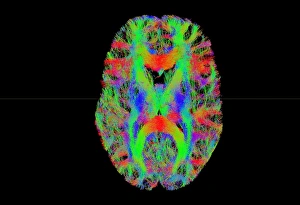
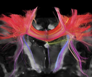
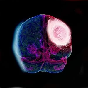
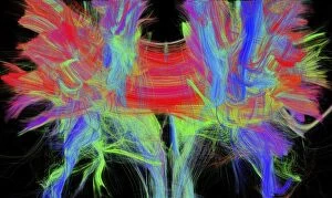
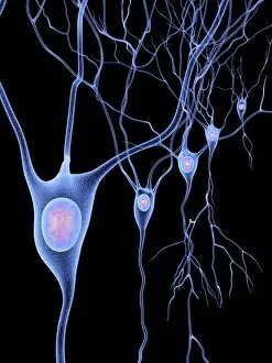
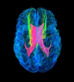
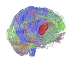
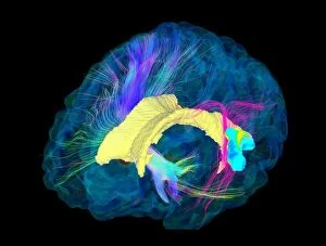
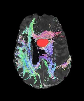
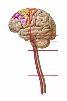
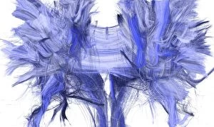
"Unraveling the Intricacies of Neural Pathways: A Journey into the Brain's Fibres" The human brain, with its complex network of neural pathways, continues to fascinate scientists and researchers. Through advanced imaging techniques such as DTI MRI scans (C017 / 7099), we can now delve deeper into understanding these intricate connections. Intriguingly, each image captured through DTI MRI reveals a unique perspective on the brain's fibres. Whether it is the mesmerizing patterns seen in DTI MRI scan C017 / 7035 or the artistic representation of brain motor cortex pathways in artwork C016 / 6532, every glimpse offers valuable insights into our cognitive functioning. However, not all discoveries are without challenges. The presence of a brain tumour can disrupt these delicate neural pathways. By utilizing fMRI and tractography techniques (C017 / 7102), experts strive to map out how this abnormality affects communication within the brain. One particular pathway that garners significant attention is the corticospinal tract. Its importance lies in facilitating voluntary movements throughout our body. With DTI MRI scan C017 / 7046 and tract density imaging (C017 / 7039), researchers aim to comprehend this crucial connection further. As we explore further into white matter using DTI MRI scans, we uncover more mysteries waiting to be unraveled. These images provide us with glimpses into both healthy brains and those affected by glioblastoma tumors – an aggressive form of cancerous growths. Through DTI modeling (C017/7060) and various scans like C017/7036, C017/7048, and C017/7055-56 showcasing glioblastoma tumors' impact on neural pathways; scientists hope to develop innovative treatment strategies for patients battling this devastating condition. With each new discovery made through these cutting-edge technologies, our understanding of neural pathways expands.