
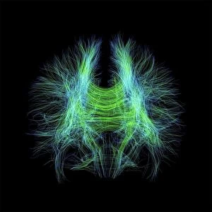
Tractography Collection
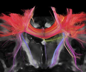
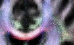
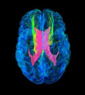
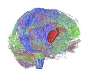
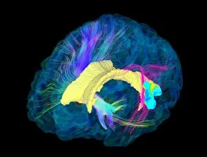
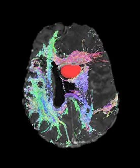
"Exploring the intricate pathways of brain fibres through tractography" Tractography, a revolutionary technique in neuroscience


All Professionally Made to Order for Quick Shipping
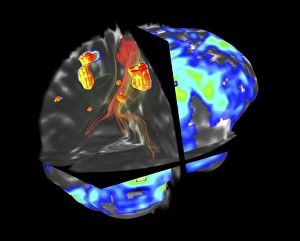
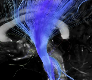
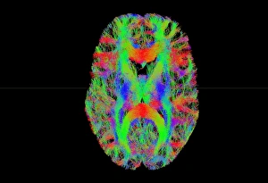
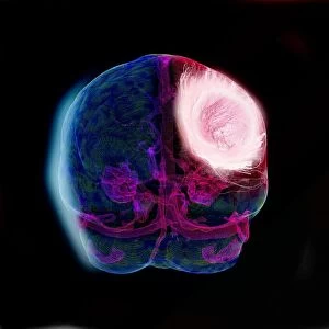
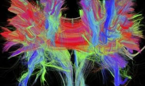
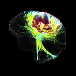
"Exploring the intricate pathways of brain fibres through tractography" Tractography, a revolutionary technique in neuroscience, allows us to delve into the complex network of brain fibres. Using advanced imaging technology such as DTI MRI scans (C017 / 7099, C017 / 7035), researchers can map out these delicate connections with unprecedented precision. In the realm of medical diagnosis and treatment, it has proven invaluable. By combining fMRI and tractography (C017 / 7102), doctors can navigate around brain tumours with greater accuracy than ever before. Specifically targeting the corticospinal tract using DTI MRI scans (C017 / 7046) enables surgeons to minimize damage during critical procedures. Moreover, tract density imaging (C017 / 7039) sheds light on the distribution and strength of brain fibres within white matter regions. This information is crucial for understanding various neurological conditions like glioblastoma brain tumours. Through DTI modelling (C017 / 7060), we gain insights into how these aggressive tumours affect fibre connectivity. Glioblastoma brain tumour cases are further examined using DTI MRI scans (C017 / 7048, C017/7055, C107/7056). These images provide vital data for treatment planning and monitoring disease progression. The corpus callosum also comes under scrutiny through DTI MRI scan analysis (C107/7044). As one of the largest white matter structures connecting both hemispheres, any abnormalities detected here could have significant implications for cognitive function. In summary, thanks to advancements in tractography techniques utilizing cutting-edge technologies like DTI MRI scans, we are unraveling the mysteries hidden within our brains' intricate web of fibres. From diagnosing diseases to guiding surgical interventions and enhancing our understanding of neural connectivity patterns – this innovative approach continues to revolutionize neuroscience research and clinical practice.