Home > Animals > Mammals > Eupleridae > Fossa
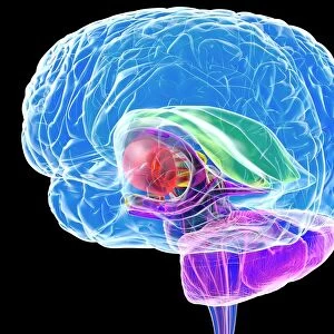
Anatomy of pituitary gland

![]()
Wall Art and Photo Gifts from Stocktrek
Anatomy of pituitary gland
Stocktrek Images specializes in Astronomy, Dinosaurs, Medical, Military Forces, Ocean Life, & Sci-Fi
Media ID 13013087
© Stocktrek Images
Anatomy Artery Biology Biomedical Illustrations Central Nervous System Cerebellum Cerebrum Connection Cross Section Cutaway View Cutout Detail Diagram Diencephalon Dissection Endocrine Glands Endocrine System Endocrinology Gland Healthcare Human Anatomy Human Body Parts Human Organs Hypothalamus Internal Organs Male Likeness Medicine Medulla Midbrain Nervous System Neurology Neuroscience Part Of Physiology Pons Posterior Sensory System Sphenoid Bone Telencephalon Adenohypophysis Anterior Pituitary Neural Tube Pituitary Gland
FEATURES IN THESE COLLECTIONS
> Animals
> Mammals
> Eupleridae
> Fossa
EDITORS COMMENTS
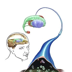
This print showcases the intricate and complex anatomy of the pituitary gland, a vital component of our endocrine system. Against a pristine white background, this close-up biomedical illustration allows us to delve into the inner workings of this remarkable organ. The image highlights various parts of the pituitary gland, including the anterior pituitary, pars tuberalis, pars distalis, hypophyseal fossa, sella turcica (within the sphenoid bone), posterior region with its pars nervosa and median eminence connected to the hypothalamus. It also provides a glimpse into its relationship with other structures such as cerebellum, cerebrum, medulla oblongata and different sections of brain like telencephalon (forebrain), diencephalon (interbrain), midbrain and pons. With meticulous detail and precision artistry in play here, this digitally generated image offers an invaluable resource for medical professionals studying neurology or endocrinology. The cutaway view reveals arteries connecting key areas while emphasizing male likeness without any human presence. As we explore this visually stunning representation of internal organs within our central nervous system's neural tube dissection becomes apparent. This artwork serves as an essential tool for understanding not only human anatomy but also how our sensory system functions at a cellular level. Without mentioning commercial use or any specific company affiliation beyond Stocktrek Images who provided it; let us appreciate this extraordinary piece that seamlessly blends science with artistic brilliance.
MADE IN THE UK
Safe Shipping with 30 Day Money Back Guarantee
FREE PERSONALISATION*
We are proud to offer a range of customisation features including Personalised Captions, Color Filters and Picture Zoom Tools
SECURE PAYMENTS
We happily accept a wide range of payment options so you can pay for the things you need in the way that is most convenient for you
* Options may vary by product and licensing agreement. Zoomed Pictures can be adjusted in the Basket.