Fraud Blocker Skip to main content

Midbrain Collection

The midbrain, a crucial part of the human brain anatomy, is depicted in this captivating caption

Background imageMidbrain Collection: Human brain anatomy, lateral view

Human brain anatomy, lateral view

Background imageMidbrain Collection: Anatomy of the human brain, lithograph, published in 1876

Anatomy of the human brain, lithograph, published in 1876
Anatomy of the human brain. Lithograph, published in 1876

Background imageMidbrain Collection: Brain motor cortex pathways, artwork C016 / 6532

Brain motor cortex pathways, artwork C016 / 6532
Brain motor cortex pathways. Artwork of a sectioned human brain, brainstem and spinal cord, showing neural pathways (red and blue) from the motor cortex of the brain

Background imageMidbrain Collection: Human head anatomy, artwork

Human head anatomy, artwork
Human head anatomy, computer artwork

Background imageMidbrain Collection: Human body showing autonomic nervous system and limbic system

Human body showing autonomic nervous system and limbic system
Side view of human body showing autonomic nervous system and limbic system within the brain. Green are parasympathetic nerves. Blue are sympathetic nerves

Background imageMidbrain Collection: Brain surface anatomy, with labels

Brain surface anatomy, with labels

Background imageMidbrain Collection: Side view of the human brain showing the functional lobes

Side view of the human brain showing the functional lobes

Background imageMidbrain Collection: Transverse section of the midbrain

Transverse section of the midbrain

Background imageMidbrain Collection: Digital cross section illustration of impulses from lower brain stem passing to inferior

Digital cross section illustration of impulses from lower brain stem passing to inferior colliculus of midbrain

Background imageMidbrain Collection: Digital illustration of highlighted areas in human brain affected by motor disorders

Digital illustration of highlighted areas in human brain affected by motor disorders

Background imageMidbrain Collection: Brain surface anatomy

Brain surface anatomy

Background imageMidbrain Collection: Pathway of a pain message via sensory nerve in injured muscle

Pathway of a pain message via sensory nerve in injured muscle, to pain gate in spinal cord to limbic system, frontal cortex and sensory cortex in the human brain

Background imageMidbrain Collection: Anatomy of pituitary gland

Anatomy of pituitary gland

Background imageMidbrain Collection: Representation of how our senses affect our thoughts

Representation of how our senses affect our thoughts

Background imageMidbrain Collection: Side view of the human head showing the functional lobes of the brain

Side view of the human head showing the functional lobes of the brain
Side view of the human head showing the functional lobes of the human brain

Background imageMidbrain Collection: The sympathetic nervous system and the organs of fight-or-flight response

The sympathetic nervous system and the organs of fight-or-flight response

Background imageMidbrain Collection: View of limbic system as seen from directly above the head

View of limbic system as seen from directly above the head

Background imageMidbrain Collection: Cross section of brain with arteries

Cross section of brain with arteries

Background imageMidbrain Collection: Human brain showing the layout of the sensory cortex

Human brain showing the layout of the sensory cortex
Coronal section through human brain showing the layout of the sensory cortex

Background imageMidbrain Collection: Endorphins released in the hypothalamus travel down the spinal cord

Endorphins released in the hypothalamus travel down the spinal cord
Serotonin released in the brain travels down the spinal cord to close the pain gates and block pain messages

Background imageMidbrain Collection: Sagittal view of human brain showing the corpus callosum

Sagittal view of human brain showing the corpus callosum

Background imageMidbrain Collection: Cutaway view of human skull showing brain details, side view

Cutaway view of human skull showing brain details, side view

Background imageMidbrain Collection: Side view of human brain showing internal carotid artery

Side view of human brain showing internal carotid artery

Background imageMidbrain Collection: X-ray view of human face, profile view

X-ray view of human face, profile view

Background imageMidbrain Collection: Detail of ateries of the human head and the trigeminal nerve

Detail of ateries of the human head and the trigeminal nerve

Background imageMidbrain Collection: Human brain anatomy, lateral view, with labels

Human brain anatomy, lateral view, with labels

Background imageMidbrain Collection: Human brain

Human brain

Background imageMidbrain Collection: Brain synapse, anatomical artwork

Brain synapse, anatomical artwork
Brain synapse. Anatomical computer artwork of a human brain with an enlargement showing the structure of a synapse (lower right) within one of the striate bodies that make up the striatum

Background imageMidbrain Collection: Striatum, anatomical artwork

Striatum, anatomical artwork
Striatum. Anatomical computer artwork of a human brain, showing the striate bodies (blue and green) in the midbrain, and the substantia nigra (black) in the brainstem

Background imageMidbrain Collection: Substantia nigra, artwork

Substantia nigra, artwork
Substantia nigra. Computer artwork showing a healthy substantia nigra (left) and degenerated substantia nigra (right) from human brains

Background imageMidbrain Collection: Brain motor cortex pathways, artwork C016 / 6533

Brain motor cortex pathways, artwork C016 / 6533
Brain motor cortex pathways. Artwork of a sectioned human brain, brainstem and spinal cord, showing neural pathways (black lines) from the motor cortex of the brain

Background imageMidbrain Collection: Brain motor cortex pathways, artwork C016 / 6531

Brain motor cortex pathways, artwork C016 / 6531
Brain motor cortex pathways. Artwork of a lateral (from the side) view of a human brain, showing neural pathways (pink lines) from the motor cortex of the brain to the brainstem and spinal cord


All Professionally Made to Order for Quick Shipping

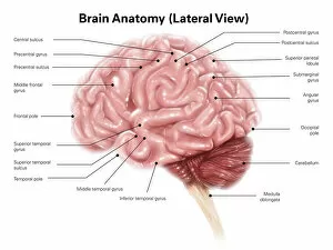
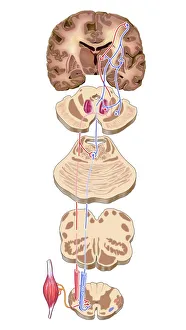
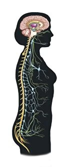
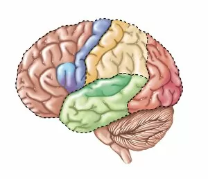
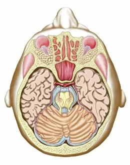
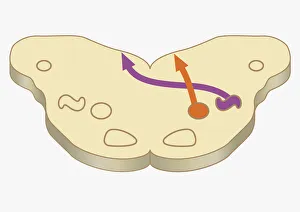
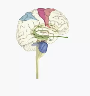
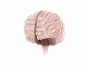
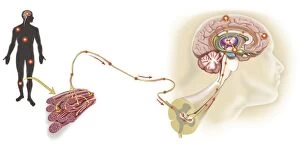
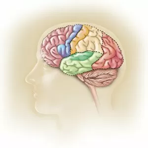
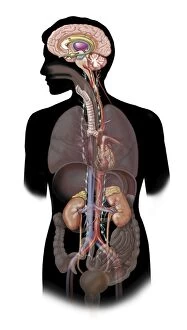
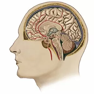
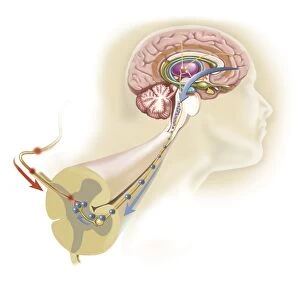
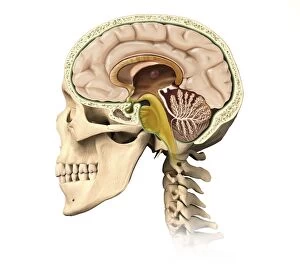
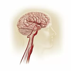
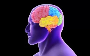
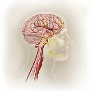
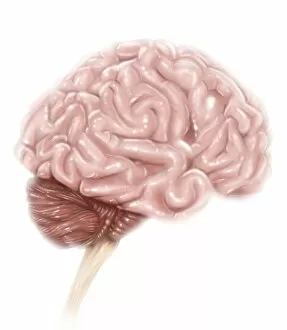
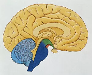
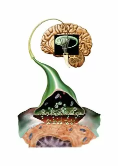
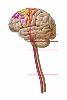
The midbrain, a crucial part of the human brain anatomy, is depicted in this captivating caption. In a lithograph published back in 1876, we get a lateral view of the intricate anatomy of the human brain. The artwork C016 / 6532 showcases the brain motor cortex pathways, highlighting its significance in our body's movement control. Moving on to another illustration, we witness the complexity of our autonomic nervous system and limbic system within the human body. This depiction emphasizes how these systems play an essential role in regulating involuntary bodily functions and emotions. A closer look at the surface anatomy of our brain reveals labeled regions that help us understand its functionality better. Additionally, a digital cross-section illustration demonstrates impulses from lower brain stem passing to inferior colliculus of midbrain—a process vital for relaying auditory information. Motor disorders affecting certain areas within our brains are highlighted through detailed digital illustrations. These visuals shed light on specific regions impacted by such disorders and emphasize their importance for proper bodily movements. Furthermore, exploring pain messages via sensory nerves originating from injured muscles unveils yet another aspect function—the pathway through which pain signals travel. Delving deeper into neuroanatomy, we encounter an exploration of pituitary gland structure—an organ responsible for hormone regulation and homeostasis maintenance within our bodies. Lastly, this captivating caption reminds us how our senses profoundly impact our thoughts—depicting how external stimuli shape and influence cognitive processes occurring within different parts of our remarkable brains.