Fraud Blocker Skip to main content

Physiology Collection

"Unlocking the Secrets of the Human Body

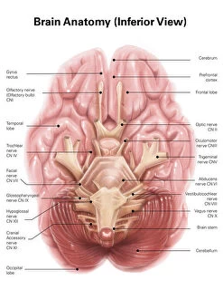
Background imagePhysiology Collection: Anatomy of human brain, inferior view

Anatomy of human brain, inferior view

Background imagePhysiology Collection: Anatomy of human knee joint

Anatomy of human knee joint

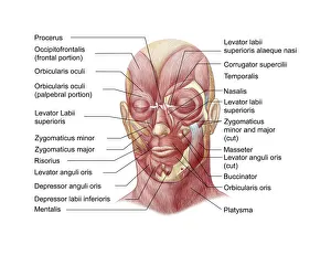
Background imagePhysiology Collection: Facial muscles of the human face (with labels)

Facial muscles of the human face (with labels)

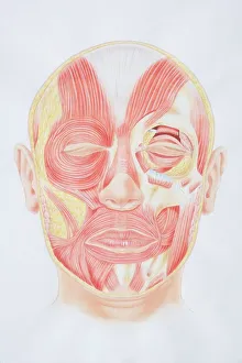
Background imagePhysiology Collection: Diagram of facial muscles, front view

Diagram of facial muscles, front view

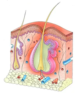
Background imagePhysiology Collection: Human hair follicle and skin

Human hair follicle and skin

Background imagePhysiology Collection: Muscular system on head

Muscular system on head
Muscular system on a head. Computer artwork showing the musculature of the side of a human males head. The muscles shown are skeletal muscles. These muscles are consciously controlled by the brain

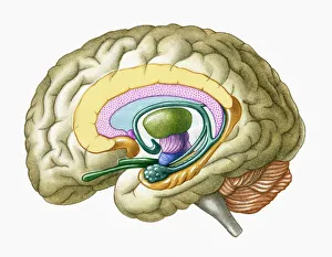
Background imagePhysiology Collection: Cross section illustration of human brain showing limbic system and primitive forebrain

Cross section illustration of human brain showing limbic system and primitive forebrain

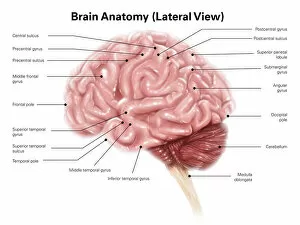
Background imagePhysiology Collection: Human brain anatomy, lateral view

Human brain anatomy, lateral view

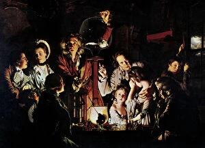
Background imagePhysiology Collection: The Airpump by Joseph Wright

The Airpump by Joseph Wright
The Air Pump by Joseph Wright (1734-1797). This artwork was painted in 1768. Its full title is An Experiment on a Bird in the Air Pump

Background imagePhysiology Collection: Cell types in the mammalian cerebellum: drawing, 1894, by the Spanish histologist Santiago Ramon y

Cell types in the mammalian cerebellum: drawing, 1894, by the Spanish histologist Santiago Ramon y Cajal (1852-1934)
NERVE CELLS, 1894. Cell types in the mammalian cerebellum: drawing, 1894, by the Spanish histologist Santiago Ramon y Cajal (1852-1934)

Background imagePhysiology Collection: Illustration of human lymphatic system

Illustration of human lymphatic system

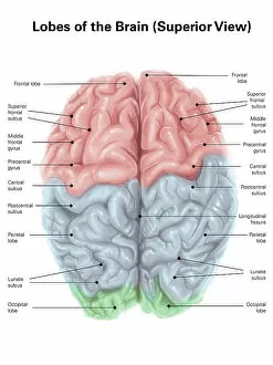
Background imagePhysiology Collection: Superior view of human brain with colored lobes and labels

Superior view of human brain with colored lobes and labels

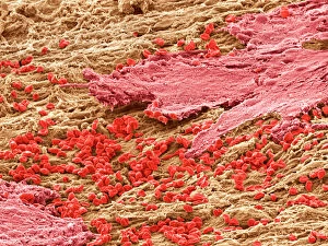
Background imagePhysiology Collection: Uterus lining during menstruation, SEM

Uterus lining during menstruation, SEM
Uterus during menstruation. Coloured scanning electron micrograph (SEM) of the lining of the uterus being shed during menstruation

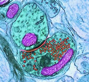
Background imagePhysiology Collection: Synapse nerve junction, TEM

Synapse nerve junction, TEM
Synapse. Coloured transmission electron micrograph (TEM) of a synapse, a junction between two nerve cells, in the brain. At a synapse an electrical signal is transmitted from one cell to the next in

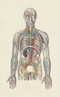
Background imagePhysiology Collection: Human circulatory system, hand-coloured engraving, published in 1861

Human circulatory system, hand-coloured engraving, published in 1861
The human circulatory system. Hand-coloured engraving, published in 1861

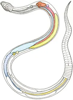
Background imagePhysiology Collection: Illustration of internal organs of a snake including heart, lung, intestines, pancreas

Illustration of internal organs of a snake including heart, lung, intestines, pancreas, kidney and testis

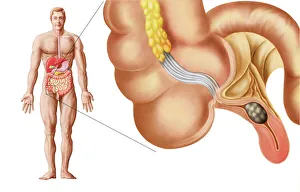
Background imagePhysiology Collection: Medical ilustration of an appendix with appendicitis

Medical ilustration of an appendix with appendicitis

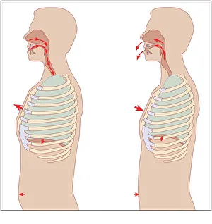
Background imagePhysiology Collection: Mechanics of respiration, diagram

Mechanics of respiration, diagram
Mechanics of respiration. Cutaway diagrams showing the mechanics of breathing in (left) and breathing out (right). Arrows show: the movement of air in and out of the nose

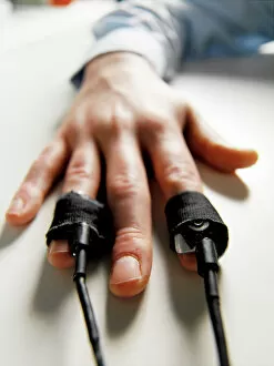
Background imagePhysiology Collection: Lie detector test

Lie detector test
MODEL RELEASED. Lie detector test. Pulse meters on a mans fingers during a lie detector, or polygraph, test. A polygraph records changes in a number of physiological processes

Background imagePhysiology Collection: Rough endoplasmic reticulum, TEM

Rough endoplasmic reticulum, TEM
Rough endoplasmic reticulum, coloured transmission electron micrograph (TEM). This section shows the rough endoplasmic reticulum (ER, folds, centre), a membranous structure that occurs in cells

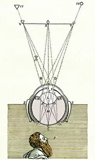
Background imagePhysiology Collection: Descartes optics theory, 17th century

Descartes optics theory, 17th century
Descartes optics theory, artwork. This is the eye diagram from Descartes book Optics (1637). Three external points are shown depicted on the retina at the back of the eye

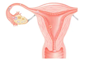
Background imagePhysiology Collection: Digital illustration showing journey of fertilized human egg

Digital illustration showing journey of fertilized human egg

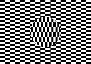
Background imagePhysiology Collection: Ouchi illusion

Ouchi illusion. The central circular area of perpendicularly orientated bars appears to move and float compared to the surrounding pattern

Background imagePhysiology Collection: Blood cells

Blood cells, computer artwork. Red blood cells (red), or erythrocytes, are biconcave disc-shaped cells that are responsible for supplying tissues with oxygen

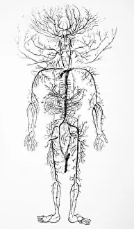
Background imagePhysiology Collection: Arterial system, 18th century

Arterial system, 18th century. Artwork of the layout and appearance of the arteries of the human circulatory system. This artwork is from Encyclopedie

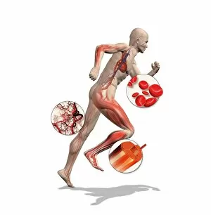
Background imagePhysiology Collection: Athlete physiology, artwork

Athlete physiology, artwork
Athlete physiology. Computer artwork of a male athlete, with enlargements to show the important physiological enhancements typical of most athletes

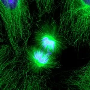
Background imagePhysiology Collection: Cell division, fluorescent micrograph

Cell division, fluorescent micrograph
Cell division. Immunofluorescent light micrograph of a human epithelial cell (centre) during the late anaphase stage of mitosis

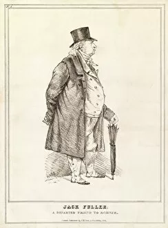
Background imagePhysiology Collection: John Fuller, English philanthropist

John Fuller, English philanthropist
John Fuller (1757-1834), English philanthropist. Fuller was born at North Stoneham, Hampshire. At the age of four he lost his father, the local clergyman, and at ten was sent to Eton College
Background imagePhysiology Collection: John Mad Jack Fuller, philanthropist

John Mad Jack Fuller, philanthropist
John Fuller (1757-1834), English philanthropist. Fuller was born at North Stoneham, Hampshire. At the age of four he lost his father, the local clergyman, and at ten was sent to Eton College
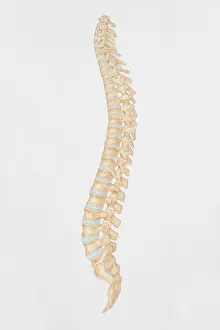
Background imagePhysiology Collection: Diagram of the human spine, side view

Diagram of the human spine, side view

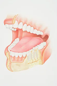
Background imagePhysiology Collection: Cross-section diagram of human mouth and jaw

Cross-section diagram of human mouth and jaw

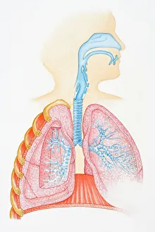
Background imagePhysiology Collection: Illustration of human respiratory system showing oral cavity, and nasal cavity, larynx, trachea

Illustration of human respiratory system showing oral cavity, and nasal cavity, larynx, trachea, bronchus, and lungs

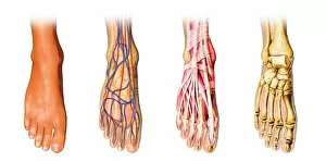
Background imagePhysiology Collection: Human foot anatomy showing skin, veins, arteries, muscles and bones

Human foot anatomy showing skin, veins, arteries, muscles and bones
Human foot anatomy showing skin, veins, arteries, muscles, and bones, cutaway view

Background imagePhysiology Collection: Hand

Hand
Antique illustration of a human hand with tendons

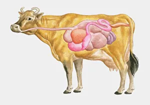
Background imagePhysiology Collection: Cross section illustration of cow digestive system

Cross section illustration of cow digestive system

Background imagePhysiology Collection: Brain motor cortex pathways, artwork C016 / 6532

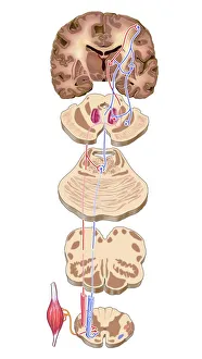
Brain motor cortex pathways, artwork C016 / 6532
Brain motor cortex pathways. Artwork of a sectioned human brain, brainstem and spinal cord, showing neural pathways (red and blue) from the motor cortex of the brain

Background imagePhysiology Collection: Blood coagulation cascade, artwork C016 / 9873

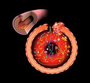
Blood coagulation cascade, artwork C016 / 9873
Blood coagulation cascade. Artwork of the biochemical cascade of blood chemicals and proteins during blood clotting (coagulation). The blood vessel and its layered wall is at upper left

Background imagePhysiology Collection: Konstantin Buteyko, Soviet doctor

Konstantin Buteyko, Soviet doctor
Konstantin Pavlovich Buteyko (1923-2003), Soviet medical doctor and researcher. In the 1950s, Buteyko, a Ukrainian, developed a theory on the effects of deep breathing (hyperventilation)

Background imagePhysiology Collection: Brain neuron

Brain neuron. Computer reconstruction of a medium spiny neuron from the basal ganglia of the brain. Neurons (nerve cells) are responsible for passing information around the central nervous system

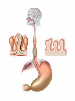
Background imagePhysiology Collection: Human digestive system, artwork

Human digestive system, artwork. At top, food from the mouth forms a bolus that is swallowed down the oesophagus. At centre, the bolus is about to enter the stomach (arrow)

Background imagePhysiology Collection: Theodor Schwann, German physiologist

Theodor Schwann, German physiologist who made many contributions to biology and coined the term metabolism

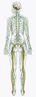
Background imagePhysiology Collection: Digital illustration of human nervous system connected to spinal cord and brain

Digital illustration of human nervous system connected to spinal cord and brain

Background imagePhysiology Collection: Digital illustration of Bocas area (purple), Wernickes area (orange)

Digital illustration of Bocas area (purple), Wernickes area (orange), Geschwinds territory (turquoise) and arcuate fasciculus (green) in human brain

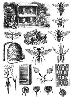
Background imagePhysiology Collection: Beekeeping

Beekeeping (or apiculture, from Latin: apis bee ) is the maintenance of honey bee colonies, commonly in hives

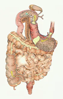
Background imagePhysiology Collection: Illustration of human digestive system, including stomach, small intestine and large intestine

Illustration of human digestive system, including stomach, small intestine and large intestine

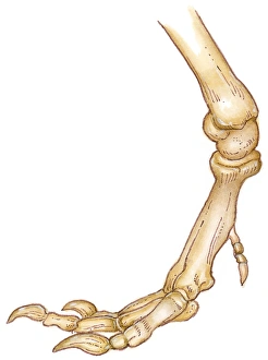
Background imagePhysiology Collection: Illustration of bones of Tyrannosaurus foot showing long toes, small first toe, or dew claw

Illustration of bones of Tyrannosaurus foot showing long toes, small first toe, or dew claw, and ankle joint

Background imagePhysiology Collection: Digital cross section illustration of taste buds and filiform papilae on tongue

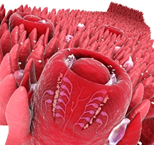
Digital cross section illustration of taste buds and filiform papilae on tongue

Background imagePhysiology Collection: Illustration of response to infection after vaccination, involving microbe, antigens, antibody, plas

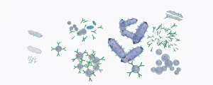
Illustration of response to infection after vaccination, involving microbe, antigens, antibody, plas


All Professionally Made to Order for Quick Shipping

"Unlocking the Secrets of the Human Body: Exploring Physiology from Head to Toe" Dive into the intricate world of human physiology with a captivating view of the anatomy of the brain, seen from an inferior perspective. Step back in time and witness Joseph Wright's masterpiece, "The Airpump, " as it symbolizes our quest to understand how our bodies function on a physiological level. Discover the hidden powerhouses behind every smile and frown with a detailed diagram showcasing facial muscles labeled for your convenience. Take a front-row seat to unravel the complexity of facial muscles as they work harmoniously together, providing us with countless expressions that define our humanity. Delve into knee joint mechanics and gain insight into how this remarkable structure allows us to walk, run, and perform various movements effortlessly. Journey beneath your skin's surface and explore the fascinating relationship between hair follicles and skin – two interconnected systems that contribute to our unique appearance. Witness synapse nerve junctions through a TEM image, offering a glimpse into one of nature's most efficient communication networks within our nervous system. Marvel at the intricacies of head muscles as they enable us to express emotions, chew food, speak articulately, or simply turn our heads towards something captivating. Uncover ancient evolutionary structures within our brains by examining cross-section illustrations depicting limbic system functions alongside primitive forebrain regions. Embark on an enlightening tour through your body's drainage system - discover how lymphatic vessels play an essential role in maintaining overall health and immunity. Explore superior views of colored lobes accompanied by labels; see how different brain regions contribute uniquely to cognition, emotion regulation & sensory processing Dissecting human brain anatomy from lateral view reveals its awe-inspiring complexity; marvel at its convoluted folds & diverse functional areas shaping who we are.