
Pituitary Gland Collection
The pituitary gland, also known as the master gland, plays a crucial role in our body's internal systems


All Professionally Made to Order for Quick Shipping
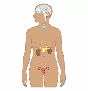
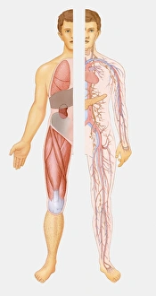
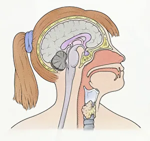
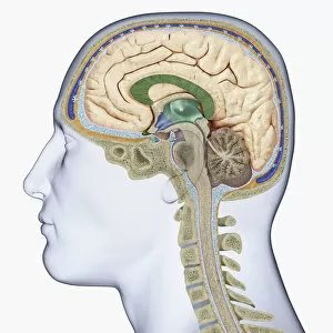
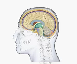
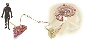
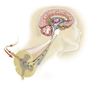
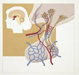
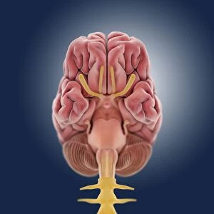
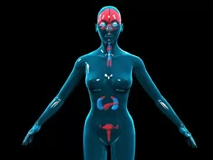
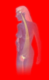
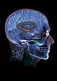
The pituitary gland, also known as the master gland, plays a crucial role in our body's internal systems, and is responsible for producing and releasing various hormones that regulate growth, metabolism, reproduction, and many other bodily functions. One of the key hormones produced by the the human growth hormone molecule. This hormone stimulates growth and development in children and helps maintain healthy tissues throughout adulthood. In an illustration of the internal systems of the human body, we can see how the pituitary gland is located at the base of the brain, and is connected to both the hypothalamus and thyroid gland through intricate pathways. A cross-section biomedical illustration shows us how these connections work in an adult female. The thyroid gland relies on signals from the pituitary gland to release its own hormones that control metabolism and energy levels. In another illustration depicting a pre-adolescent girl's brain, we can observe how closely linked her thyroid and they are to her brain. These connections highlight their vital roles in regulating hormonal balance during critical stages of development. A digital cross-section illustration reveals more details about this complex relationship between the hypothalamus (a region within our brain) and pituitary gland. Together they form a powerful duo responsible for coordinating numerous bodily functions. Moving on to a profile view of our head, separate areas of our brain are highlighted using different colors. Among them stands out our precious pituitary gland - a small but mighty organ with immense influence over our overall well-being. However, sometimes things can go awry within this delicate system. A digital illustration showcases a head profile with a highlighted section revealing a pituitary tumor present in someone's brain. Such tumors can disrupt normal hormone production leading to various health complications. Zooming back into just highlighting specific regions within our head profile image; here we focus on thalamus (green), hypothalamus (blue), and the pituitary gland (also blue).