
Biomedical Illustrations Collection
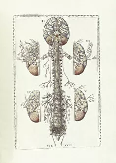
"Exploring the Intricacies of Biomedical Illustrations: A Journey into the Human Body" Anatomy of human brain


All Professionally Made to Order for Quick Shipping
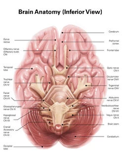
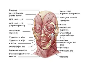
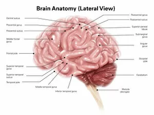
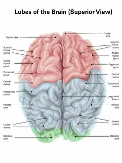
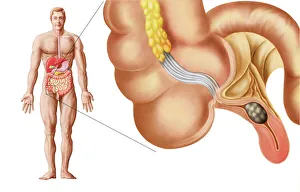
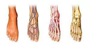
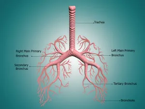
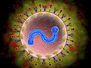
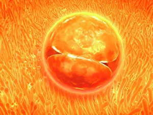
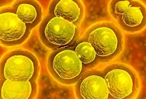
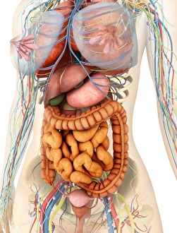
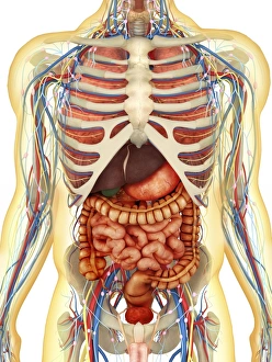
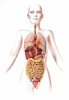
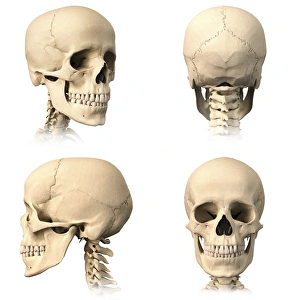
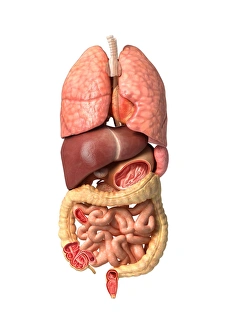
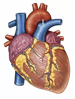
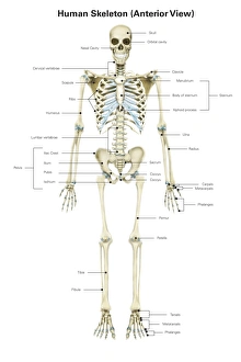
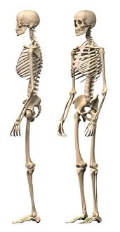
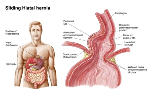
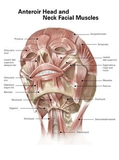
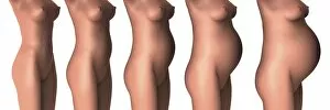
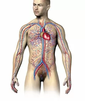
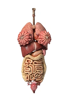
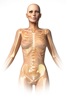
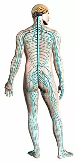
"Exploring the Intricacies of Biomedical Illustrations: A Journey into the Human Body" Anatomy of human brain, inferior view: Delve into the complex network of neurons and discover the hidden wonders within our minds. Facial muscles of the human face (with labels): Uncover the intricate web of muscles that enable us to express emotions and communicate nonverbally. Superior view of human brain with colored lobes and labels: Take a colorful tour through different regions of our brain, each responsible for unique functions that shape who we are. Human brain anatomy, lateral view: Witness the elegance and complexity of our most vital organ from a side perspective, unveiling its remarkable structures and pathways. Medical illustration of an appendix with appendicitis: Explore a visual representation showcasing how this small organ can cause significant discomfort when afflicted by inflammation. The human body with superimposed colored plates by Julien Bougle: Experience an artistic fusion where vibrant hues bring life to anatomical structures, revealing their interconnectedness in stunning detail. Human foot anatomy showing skin, veins, arteries, muscles, and bones: Step into a comprehensive depiction highlighting every aspect that makes up our foundation - from delicate skin to robust bones - enabling us to walk through life's journey. Anatomy of bronchus and bronchial tubes: Dive deep into respiratory system intricacies as you unravel how air travels through branching passageways within your lungs. Microscopic view of human respiratory syncytial virus: Peer through a microscope lens at this microscopic menace wreaking havoc on our respiratory systems – gaining insight into its structure for potential treatments or prevention strategies. Embryo development 24-36 hours after fertilization: Witness nature's miracle unfold as cells multiply rapidly during early embryonic stages – setting in motion the formation of new life itself. Conceptual image of human skull and spinal cord.