Home > Popular Themes > Human Body
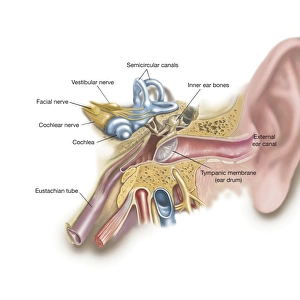
Anatomy of the cochlear duct in the human ear

![]()
Wall Art and Photo Gifts from Stocktrek
Anatomy of the cochlear duct in the human ear
Stocktrek Images specializes in Astronomy, Dinosaurs, Medical, Military Forces, Ocean Life, & Sci-Fi
Media ID 13012773
© Stocktrek Images
Acoustic Anatomy Audio Auditory Auditory System Aural Auricle Basilar Membrane Biology Biomedical Illustrations Cell Cochlea Cochlear Duct Cross Section Cutaway View Detail Diagram Dissection Ear Canal Ear Drum Endolymph Eustachian Tube Gland Health Healthcare Hearing Human Anatomy Human Body Human Body Parts Human Ear Human Glands Human Organs Human Tissue Incus Inner Ear Internal Organs Listening Magnification Malleus Meatus Medical Medicine Membrane Membranous Labyrinth Middle Ear Nerve Organ Organ Of Corti Ossicle Outer Ear Physiology Pinna Scala Tympani Scala Vestibuli Scarpas Ganglion Sensory System Sound Spiral Spiral Ganglion Stapes Structure Text Tympanic Membrane Utricle Vestibular Nerves Vestibular System Western Script Modiolus Semicircular Canal
FEATURES IN THESE COLLECTIONS
EDITORS COMMENTS
This visually stunning print showcases the intricate anatomy of the cochlear duct in the human ear. With a white background, this digitally generated illustration brings to life the complex structure and function of this vital organ within our auditory system. The horizontal composition allows for a detailed close-up view, revealing every cell and cross section with remarkable clarity. The image highlights various components such as the ear canal, cochlea, ossicles, tympanic membrane, and nerve pathways that contribute to our sense of hearing. It also includes important elements like the organ of Corti, vestibular nerves, basilar membrane, Reissner's membrane, and endolymph glands. Through meticulous dissection and magnification techniques employed in biomedical illustrations like this one by Stocktrek Images, we gain a deeper understanding of how sound waves are transformed into electrical signals that our brain can interpret. This artwork serves as an invaluable resource for medical professionals in fields such as otology or audiology. Its scientific accuracy combined with artistic flair makes it equally captivating for anyone interested in exploring the wonders of human biology. Without mentioning commercial use or any specific company affiliation, this print is a testament to Stocktrek Images' commitment to providing high-quality medical imagery that educates and inspires viewers worldwide.
MADE IN THE UK
Safe Shipping with 30 Day Money Back Guarantee
FREE PERSONALISATION*
We are proud to offer a range of customisation features including Personalised Captions, Color Filters and Picture Zoom Tools
SECURE PAYMENTS
We happily accept a wide range of payment options so you can pay for the things you need in the way that is most convenient for you
* Options may vary by product and licensing agreement. Zoomed Pictures can be adjusted in the Basket.