Fraud Blocker Skip to main content

Auditory Collection

"Exploring the Wonders of Auditory Perception

Background imageAuditory Collection: Diagram of inner ear showing auditory canal, eardrum, semicircular canals, cochlea, cochlea nerve

Diagram of inner ear showing auditory canal, eardrum, semicircular canals, cochlea, cochlea nerve, eustachian tube

Background imageAuditory Collection: Human ear anatomy, artwork

Human ear anatomy, artwork
Human ear anatomy, computer artwork

Background imageAuditory Collection: Paris: Auditory of the Church of the Invalides (b / w photo)

Paris: Auditory of the Church of the Invalides (b / w photo)
6007940 Paris: Auditory of the Church of the Invalides (b/w photo) by French Photographer, (19th century); Private Collection; (add.info.: Auditory of the Church of the Invalides)

Background imageAuditory Collection: Allegory of human hearing and music, from Utriusque Cosmi Historia Tractatus

Allegory of human hearing and music, from Utriusque Cosmi Historia Tractatus
CHT360253 Allegory of human hearing and music, from Utriusque Cosmi Historia Tractatus Secundus de naturae Simia seu Tecnica Macrocosmi, published in Frankfurt, 1617-24 (engraving) by German School

Background imageAuditory Collection: Section of One of the Free Churches of Scotland, showing the Mode of Ventilating the Auditory

Section of One of the Free Churches of Scotland, showing the Mode of Ventilating the Auditory (engraving)
1594081 Section of One of the Free Churches of Scotland, showing the Mode of Ventilating the Auditory (engraving) by English School

Background imageAuditory Collection: anatomy, auditory, auditory canal, auditory ossicles, auditory system, black background

anatomy, auditory, auditory canal, auditory ossicles, auditory system, black background
anatomy, auditory, auditory canal, auditory ossicles, auditory syste, Medical Image Collection, 87396781

Background imageAuditory Collection: Anatomy of the cochlear duct in the human ear

Anatomy of the cochlear duct in the human ear

Background imageAuditory Collection: Anatomy of human ear

Anatomy of human ear

Background imageAuditory Collection: External auditory canal of human ear (with labels)

External auditory canal of human ear (with labels)

Background imageAuditory Collection: Anatomy of inner ear and sinuses

Anatomy of inner ear and sinuses

Background imageAuditory Collection: Cutaway diagram of human ear

Cutaway diagram of human ear

Background imageAuditory Collection: Human ear C013 / 7319

Human ear C013 / 7319
MODEL RELEASED. Human ear. Close-up of the ear of a 24-year-old man. The visible structure of the ear consists of the folds and curves of the pinna

Background imageAuditory Collection: Pipistrelle bat ear, SEM

Pipistrelle bat ear, SEM
Pipistrelle bat ear. Coloured scanning electron micrograph (SEM) of the ear of a pipistrelle bat (Pipistrellus pipistrellus)

Background imageAuditory Collection: Human ear anatomy

Human ear anatomy. Computer artwork of the structure of the human ear, showing the outer ear (left), middle ear and inner ear (right)

Background imageAuditory Collection: Medical illustration of endolymph in the membranous labyrinth of the inner ear

Medical illustration of endolymph in the membranous labyrinth of the inner ear
Medical ilustration showing endolymph in the membranous labyrinth of the inner ear

Background imageAuditory Collection: Anatomy of the organ of Corti, part of the cochlea of the inner ear

Anatomy of the organ of Corti, part of the cochlea of the inner ear

Background imageAuditory Collection: Interior detail of the cochlea

Interior detail of the cochlea

Background imageAuditory Collection: Anatomy of human ear, membranous labyrinth

Anatomy of human ear, membranous labyrinth

Background imageAuditory Collection: Microscopic view of the cochlea of the inner ear

Microscopic view of the cochlea of the inner ear. The cochlea contains the spiral organ of Corti, which is the receptor organ for hearing

Background imageAuditory Collection: Cross section of human ear

Cross section of human ear

Background imageAuditory Collection: Cochlea detail with vestibulocochlear nerve

Cochlea detail with vestibulocochlear nerve

Background imageAuditory Collection: External auditory canal of human ear

External auditory canal of human ear

Background imageAuditory Collection: KCNQ ion channel protein structure

KCNQ ion channel protein structure. Molecular model showing the protein structure of an ion channel domain. Ion channels are membrane-spanning proteins that form a pathway for the movement of

Background imageAuditory Collection: Structure of the cochlea, artwork

Structure of the cochlea, artwork
Structure of the cochlea. Computer artwork of a section through the organ of Corti, the auditory sense organ that lines the spiral of the cochlea in the inner ear

Background imageAuditory Collection: Hearing centre, artwork

Hearing centre, artwork
Hearing centre. Computer artwork showing the area of the brains temporal lobe that is associated with hearing (red). This area receives auditory input from the cochlea (coiled)

Background imageAuditory Collection: Cochlear implant, artwork C016 / 7683

Cochlear implant, artwork C016 / 7683
Cochlear implant. Computer artwork of a section through the outer (left) and inner (right) ear showing a cochlear implant (top left), a prosthetic hearing device

Background imageAuditory Collection: Fish inner ear, artwork

Fish inner ear, artwork
Fish inner ear. Computer artwork showing the position and structure (inset) of the inner ear of a fish

Background imageAuditory Collection: Eustachian tube anatomy, artwork

Eustachian tube anatomy, artwork
Eustachian tube anatomy. Artwork of the anatomy of the human Eustachian tube in its normal closed state (main artwork) and in an open state (inset, upper right)

Background imageAuditory Collection: Inner ear hair cells, SEM C014 / 4845

Inner ear hair cells, SEM C014 / 4845
Inner ear hair cells. Coloured scanning electron micrograph (SEM) showing the sensory hair cells (white) found in the Organ of Corti in the cochlea of the inner ear

Background imageAuditory Collection: Inner ear hair cells, SEM C014 / 4852

Inner ear hair cells, SEM C014 / 4852
Inner ear hair cells. Coloured scanning electron micrograph (SEM) showing the sensory hair cells (blue) found in the Organ of Corti in the cochlea of the inner ear

Background imageAuditory Collection: Inner ear hair cells, SEM C014 / 4851

Inner ear hair cells, SEM C014 / 4851
Inner ear hair cells. Coloured scanning electron micrograph (SEM) showing the sensory hair cells (white) found in the Organ of Corti in the cochlea of the inner ear

Background imageAuditory Collection: Inner ear hair cells, SEM C014 / 4844

Inner ear hair cells, SEM C014 / 4844
Inner ear hair cells. Coloured scanning electron micrograph (SEM) showing the sensory hair cells (blue) found in the Organ of Corti in the cochlea of the inner ear

Background imageAuditory Collection: Hearing protection, conceptual artwork

Hearing protection, conceptual artwork
Hearing protection. Conceptual artwork of a shield covering the ear of a human head surrounded by loud noise (concentric circles)

Background imageAuditory Collection: Mental disorder, conceptual artwork

Mental disorder, conceptual artwork
Mental disorder. Conceptual artwork showing unwelcome involuntary thoughts (red arrows) invading a persons mind, together with images or unpleasant ideas (top) that may become obsessions

Background imageAuditory Collection: Hearing damage, conceptual artwork

Hearing damage, conceptual artwork
Hearing damage. Conceptual artwork of a man with distorted sound waves (green) entering his ears, with a volume-indicating dial (volume unit meter) in place of his mouth

Background imageAuditory Collection: Human ears

Human ears

Background imageAuditory Collection: Earphone in an ear

Earphone in an ear
Earphone in place in an ear

Background imageAuditory Collection: Cochlear implant, artwork

Cochlear implant, artwork
Cochlear implant. Artwork of a section through the outer and inner ear showing a cochlear implant, a prosthetic hearing device

Background imageAuditory Collection: Human ear

Human ear, computer artwork

Background imageAuditory Collection: Tinnitus

Tinnitus, conceptual computer artwork. Tinnitus is experienced as ringing in the ears. It is a symptom of ear injury or hearing loss

Background imageAuditory Collection: Middle ear bone, SEM

Middle ear bone, SEM
Middle ear bone. Coloured scanning electron micrograph (SEM) of the human middle ear, showing the stapes (ring-shaped). The stapes is one of three bones (known as the ossicles)

Background imageAuditory Collection: Sensory hair cell in the ear, artwork

Sensory hair cell in the ear, artwork
Sensory hair cell in the ear. Artwork showing a section through part of the cochlea, the inner ears auditory sense organ. The hair-like cells (grey) at upper centre are stereocilia

Background imageAuditory Collection: Internal ear, artwork

Internal ear, artwork
Internal ear. Artwork of a female face showing the structure of the internal ear. The ear canal leads from the external ear (pinna) to the eardrum (tympanic membrane)

Background imageAuditory Collection: Perforated eardrum, artwork

Perforated eardrum, artwork
Perforated eardrum. Artwork of a perforated (torn) eardrum (right), or tympanic membrane, and a normal eardrum (left), as seen from the outer ear

Background imageAuditory Collection: Human ear, artwork

Human ear, artwork
Human ear. Artwork of the pinna, the external part of the ear

Background imageAuditory Collection: Ear, artwork

Ear, artwork
Ear. Computer artwork of the middle and inner ear. The middle ear includes the ear bones (ossicles, green), the ear canal (auditory canal, orange) and the ear drum (tympanic membrane, yellow)

Background imageAuditory Collection: Tinnitus, conceptual image

Tinnitus, conceptual image. Computer artwork of a bell ringing in a persons ear, representing experiencing tinnitus. Tinnitus is as ringing in the ears often caused as a symptom of ear injury or


All Professionally Made to Order for Quick Shipping

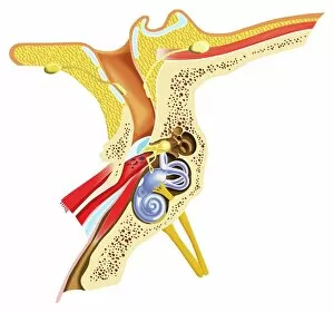
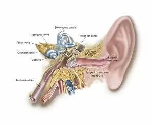
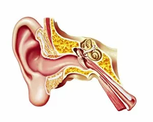
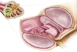
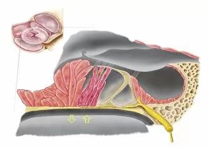
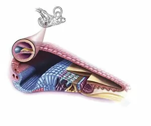
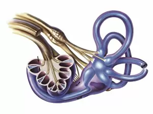
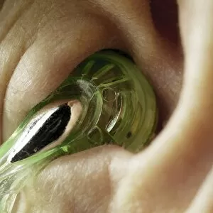
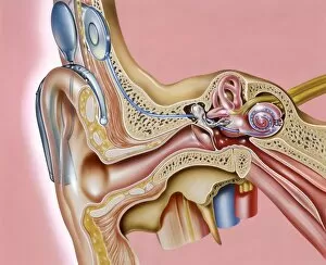
"Exploring the Wonders of Auditory Perception: From Anatomy to Music" Discovering the intricate world perception is like unraveling a captivating symphony within our very own bodies. Through scientific illustrations, we can delve into the fascinating anatomy that enables us to experience sound in all its glory. Intriguing diagrams showcase the inner ear's complexity, with its auditory canal leading to the eardrum and connecting to vital components such as semicircular canals, cochlea, cochlear nerve, and even the eustachian tube. These illustrations provide a visual gateway into understanding how our ears capture and process sound waves. Transporting ourselves from anatomical depictions to historical landmarks, we encounter Paris' magnificent Auditory of the Church of Invalides. A black-and-white photograph captures this architectural marvel where echoes of prayers once reverberated through its walls – an auditory testament to faith and devotion. Venturing further into history's embrace, an allegorical artwork portrays human hearing intertwined with music itself. This enchanting portrayal reminds us that music holds a profound connection with our ability to perceive sounds; it transcends language barriers and speaks directly to our souls. Shifting gears towards practicality, an engraving reveals how one Free Church in Scotland employed innovative ventilation techniques within their auditoriums. By ensuring fresh air circulation during gatherings or sermons, they prioritized both comfort and optimal acoustic experiences for their congregations. Returning back to anatomy's realm, detailed images shed light on facial nerves intricately connected with our auditory system – emphasizing how these two senses intertwine harmoniously within us. Additionally, we explore cross-sections unveiling the wonders housed within the cochlear duct while also examining labels guiding us through external auditory canals. The journey concludes by embracing comprehensive views encompassing inner ear structures alongside sinuses – reminding us of nature's remarkable design where multiple systems coexist seamlessly within our bodies.