Home > Popular Themes > Human Body
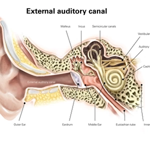
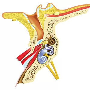
Anatomy of human ear

![]()
Wall Art and Photo Gifts from Stocktrek
Anatomy of human ear
Stocktrek Images specializes in Astronomy, Dinosaurs, Medical, Military Forces, Ocean Life, & Sci-Fi
Media ID 13011823
© TriFocal Communications/Stocktrek Images
Acoustic Anatomy Audio Auditory Auditory System Aural Auricle Biology Biomedical Illustrations Cochlea Cross Section Cutaway View Detail Diagram Dissection Ear Canal Ear Drum Eustachian Tube Healthcare Healthy Hearing Human Anatomy Human Body Human Body Parts Human Ear Human Organs Human Tissue Incus Inner Ear Internal Organs Listening Lobe Malleus Meatus Medical Medicine Membrane Middle Ear Nerve Organ Organ Of Corti Ossicle Outer Ear Physiology Pinna Scarpas Ganglion Sensory System Sound Stapes Structure Text Tympanic Membrane Utricle Vestibular System Western Script Labyrinth
FEATURES IN THESE COLLECTIONS
EDITORS COMMENTS
This print titled "Anatomy of Human Ear" offers a visually stunning depiction of the intricate inner workings of our auditory system. The horizontal, digitally generated image showcases a detailed cross-section of the ear, combining biomedical illustrations and artistic elements to create an engaging piece of artwork. Set against a clean white background, the vibrant colors and precise details bring this scientific illustration to life. From Scarpas ganglion to the cochlea, every component is meticulously portrayed, providing a comprehensive understanding of this vital sensory organ. The composition highlights various sections such as the outer ear with its pinna and ear canal, leading into the middle ear where ossicles like malleus, incus, and stapes transmit sound vibrations. Moving deeper into the inner ear reveals structures like the vestibular system responsible for balance and equilibrium. With its cutaway view and dissection-like presentation, this print invites viewers to explore human anatomy in all its complexity. It serves as a testament to both medical science's advancements and TriFocal Communications' expertise in creating educational visuals that bridge artistry with scientific accuracy. Whether you are fascinated by biology or simply intrigued by how we perceive sound, this image provides an insightful glimpse into one of our most remarkable organs – showcasing not only its structure but also celebrating its crucial role in our overall well-being.
MADE IN THE UK
Safe Shipping with 30 Day Money Back Guarantee
FREE PERSONALISATION*
We are proud to offer a range of customisation features including Personalised Captions, Color Filters and Picture Zoom Tools
SECURE PAYMENTS
We happily accept a wide range of payment options so you can pay for the things you need in the way that is most convenient for you
* Options may vary by product and licensing agreement. Zoomed Pictures can be adjusted in the Basket.