Fraud Blocker Skip to main content

Tympanic Membrane Collection

The tympanic membrane, also known as the eardrum, is a vital component of the human ear

Background imageTympanic Membrane Collection: Details of the anatomy of the human ear, showing cochlea, stapes, incus, malleus

Details of the anatomy of the human ear, showing cochlea, stapes, incus, malleus, semicircular canal, etc
FLO4611637 Details of the anatomy of the human ear, showing cochlea, stapes, incus, malleus, semicircular canal, etc. Copperplate engraving by Robert Benard after an illustration by Guichard Joseph

Background imageTympanic Membrane Collection: Anatomy of the cochlear duct in the human ear

Anatomy of the cochlear duct in the human ear

Background imageTympanic Membrane Collection: Anatomy of human ear

Anatomy of human ear

Background imageTympanic Membrane Collection: External auditory canal of human ear (with labels)

External auditory canal of human ear (with labels)

Background imageTympanic Membrane Collection: Cross section illustration of Common Earwig (Forficula auricularia) in auditory canal of ear

Cross section illustration of Common Earwig (Forficula auricularia) in auditory canal of ear, touching tympanic membrane with antennae

Background imageTympanic Membrane Collection: Anatomy of inner ear and sinuses

Anatomy of inner ear and sinuses

Background imageTympanic Membrane Collection: Cutaway diagram of human ear

Cutaway diagram of human ear

Background imageTympanic Membrane Collection: Human ear anatomy

Human ear anatomy. Computer artwork of the structure of the human ear, showing the outer ear (left), middle ear and inner ear (right)

Background imageTympanic Membrane Collection: Cross section biomedical illustration of human ear

Cross section biomedical illustration of human ear

Background imageTympanic Membrane Collection: Cross section biomedical illustration of grommet inserted in eardrum (tympanic membrane)

Cross section biomedical illustration of grommet inserted in eardrum (tympanic membrane)

Background imageTympanic Membrane Collection: Cross section biomedical illustration of internal components of the ear

Cross section biomedical illustration of internal components of the ear

Background imageTympanic Membrane Collection: Cross section biomedical illustration of the anatomy of the ear

Cross section biomedical illustration of the anatomy of the ear

Background imageTympanic Membrane Collection: Interior detail of the cochlea

Interior detail of the cochlea

Background imageTympanic Membrane Collection: Anatomy of human ear, membranous labyrinth

Anatomy of human ear, membranous labyrinth

Background imageTympanic Membrane Collection: Cross section of human ear

Cross section of human ear

Background imageTympanic Membrane Collection: Cochlea detail with vestibulocochlear nerve

Cochlea detail with vestibulocochlear nerve

Background imageTympanic Membrane Collection: External auditory canal of human ear

External auditory canal of human ear

Background imageTympanic Membrane Collection: Inner ear anatomy C018 / 6400

Inner ear anatomy C018 / 6400
Inner ear anatomy. Computer artwork of a section through the inner part of a human ear, the organ of hearing and balance. The eardrum transmits sounds, as vibrations

Background imageTympanic Membrane Collection: Inner ear anatomy C018 / 6379

Inner ear anatomy C018 / 6379
Inner ear anatomy. Computer artwork of a section through the inner part of a human ear, the organ of hearing and balance. The eardrum transmits sounds, as vibrations

Background imageTympanic Membrane Collection: Inner ear anatomy C018 / 6393

Inner ear anatomy C018 / 6393
Inner ear anatomy. Computer artwork of a section through the inner part of a human ear, the organ of hearing and balance. The eardrum transmits sounds, as vibrations

Background imageTympanic Membrane Collection: Inner ear anatomy C018 / 6387

Inner ear anatomy C018 / 6387
Inner ear anatomy. Computer artwork of a section through the inner part of a human ear, the organ of hearing and balance. The eardrum transmits sounds, as vibrations

Background imageTympanic Membrane Collection: Inner ear anatomy C018 / 6397

Inner ear anatomy C018 / 6397
Inner ear anatomy. Computer artwork of a section through the inner part of a human ear, the organ of hearing and balance. The eardrum transmits sounds, as vibrations

Background imageTympanic Membrane Collection: Inner ear anatomy C018 / 6402

Inner ear anatomy C018 / 6402
Inner ear anatomy. Computer artwork of a section through the inner part of a human ear, the organ of hearing and balance. The eardrum transmits sounds, as vibrations

Background imageTympanic Membrane Collection: Eardrum, SEM

Eardrum, SEM
Eardrum. Coloured scanning electron micrograph (SEM) of an eardrum (red). The eardrum, or tympanic membrane, is located in the middle ear

Background imageTympanic Membrane Collection: Ear anatomy

Ear anatomy. Historical anatomical artwork of a human ear. The main diagram (centre) shows the outer ear (pinna, left) and the internal structure of the ear (moving left-right): the ear canal

Background imageTympanic Membrane Collection: Inner ear anatomy, artwork

Inner ear anatomy, artwork
Inner ear anatomy. Artwork showing the anatomy of the inner human ear, the organ of hearing and balance. The ear canal (bottom) leads from the pinna (not seen), the visible part of the ear

Background imageTympanic Membrane Collection: Internal ear, artwork

Internal ear, artwork
Internal ear. Artwork of a female face showing the structure of the internal ear. The ear canal leads from the external ear (pinna) to the eardrum (tympanic membrane)

Background imageTympanic Membrane Collection: Perforated eardrum, artwork

Perforated eardrum, artwork
Perforated eardrum. Artwork of a perforated (torn) eardrum (right), or tympanic membrane, and a normal eardrum (left), as seen from the outer ear

Background imageTympanic Membrane Collection: Ear, artwork

Ear, artwork
Ear. Computer artwork of the middle and inner ear. The middle ear includes the ear bones (ossicles, green), the ear canal (auditory canal, orange) and the ear drum (tympanic membrane, yellow)

Background imageTympanic Membrane Collection: Dwarf American Toad - United States: Arizona, Illinois, Indiana, Kentucky, Missouri

Dwarf American Toad - United States: Arizona, Illinois, Indiana, Kentucky, Missouri
PM-10476 Dwarf American Toad United States: Arizona, Illinois, Indiana, Kentucky, Missouri. Bufo americanus charlesmithi Pat Morris Please note that prints are for personal display purposes only


All Professionally Made to Order for Quick Shipping

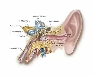
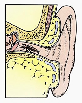
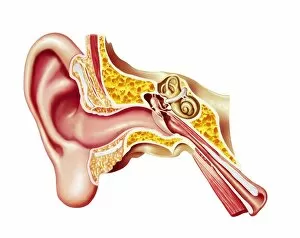
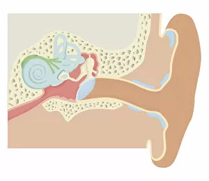
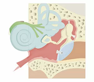
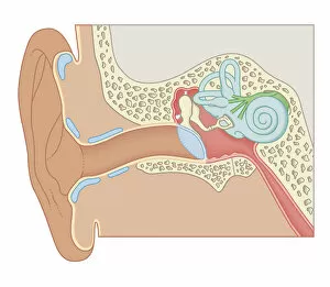
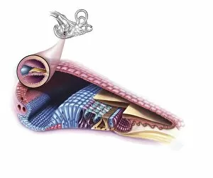
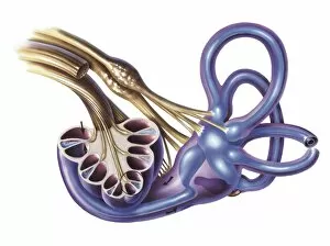
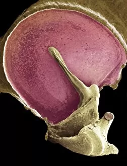
The tympanic membrane, also known as the eardrum, is a vital component of the human ear. Located at the end of the external auditory canal, it serves as a barrier between the outer and middle ear. This thin, translucent structure plays a crucial role in transmitting sound waves from the environment to the inner ear. In an intricate cross-section illustration of the common earwig (Forficula auricularia), we can observe its antennae delicately touching the tympanic membrane within the auditory canal. This interaction highlights how insects utilize their sensory organs to perceive sounds in their surroundings. Understanding the anatomy of our ears is essential for comprehending how they function. The cochlear duct, situated within this complex system, forms part of our inner ear and sinuses. A cutaway diagram provides insight into these internal components and their interconnectedness. Biomedical illustrations further demonstrate various aspects related to this fascinating organ. One such depiction showcases a grommet inserted into an eardrum affected by certain conditions or medical interventions. Another cross-sectional view emphasizes both external and internal structures present in our ears. Studying these biomedical illustrations aids researchers and healthcare professionals in diagnosing and treating disorders that affect hearing or balance. By unraveling intricacies like those found within tympanic membranes, scientists continue advancing knowledge about our remarkable auditory system.