Fraud Blocker Skip to main content

Anatomy Collection

"Unveiling the Intricacies of Anatomy: From Sensory Homunculus to Motor Homunculus" Step into the fascinating world of anatomy, where every detail tells a story

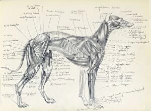
Background imageAnatomy Collection: Anatomy of a greyhound

Anatomy of a greyhound
Anatomical drawing of the muscles of a greyhound, precisely labelled

Background imageAnatomy Collection: Austalian estrildid finches

Austalian estrildid finches
An illustration by M. W. Woodcock of some Australian estrildids, published in Derek Goodwins Estrildid Finches of the World (1982). Page 96, Plate 2

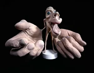
Background imageAnatomy Collection: Sensory homunculus

Sensory homunculus
This model shows what a mans body would look like if each part grew in proportion to the area of the cortex of the brain concerned with its sensory perception

Background imageAnatomy Collection: Human eye, illustration

Human eye, illustration
Eye anatomy. Cutaway illustration passing through a human eye, showing its internal anatomy and structure. The front of the eye is at right, and the structures here include the cornea

Background imageAnatomy Collection: Normal anatomy of the eye in cross section

Normal anatomy of the eye in cross section
Horizontal, Vision, Detail, Anatomy, Diagram, Muscle, Illustration, Lens, Iris, Medical, Eyeball, Biology, Lateral, Cut Out, White Background, Eye, Artwork, Cross Section, Color Image, Close Up

Background imageAnatomy Collection: Pandion haliaetus, osprey

Pandion haliaetus, osprey
Plate 15 from William MacGillivrays Watercolour drawings of British Animals (1831-1841)

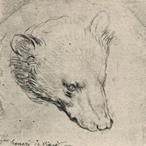
Background imageAnatomy Collection: Head of a Bear, c1480 (1945). Artist: Leonardo da Vinci

Head of a Bear, c1480 (1945). Artist: Leonardo da Vinci
Head of a Bear, c1480 (1945). From The Drawings of Leonardo da Vinci. [Reynal & Hitchcock, New York, 1945]

Background imageAnatomy Collection: Sensory homunculus

Sensory homunculus
This model shows what a mans body would look like if each part grew in proportion to the area of the cortex of the brain concerned with its sensory perception

Background imageAnatomy Collection: Skeleton of a greyhound

Skeleton of a greyhound
Detailed anatomical drawing by Raymond Sheppard in pen and ink of the skeleton of a greyhound, with bones fully labelled

Background imageAnatomy Collection: Motor homunculus

Motor homunculus
This model shows what a mans body would look like if each part grew in proportion to the area of the cortex of the brain concerned with its movement

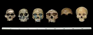
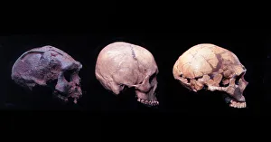
Background imageAnatomy Collection: Hominid crania

Hominid crania
L to R: Australopithecus africanus; Homo rudolfensis; H.erectus; H. heildebergensis; H. neanderthalensis and H. sapiens. Arranged in chronological order these specimens (casts)

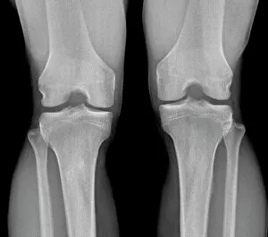
Background imageAnatomy Collection: Normal knees, X-ray

Normal knees, X-ray
Normal knees. Frontal X-ray of the flexed knees of a 24 year old patient

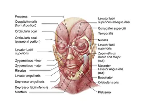
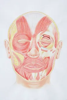
Background imageAnatomy Collection: Diagram of facial muscles, front view

Diagram of facial muscles, front view

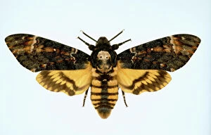
Background imageAnatomy Collection: Acherontia atropos, death s-head hawk-moth

Acherontia atropos, death s-head hawk-moth
A mounted specimen of the death s-head hawk-moth, which takes its name from the skull-like image on its thorax. Specimen from the Natural History Museum, London

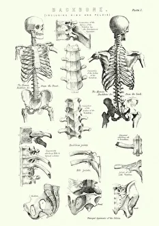
Background imageAnatomy Collection: Human Anatomy - Backbone including Ribs and Pelvis

Human Anatomy - Backbone including Ribs and Pelvis
Vintage engraving of the human Backbone including Ribs and Pelvis. 19th Century

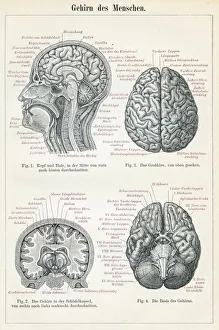
Background imageAnatomy Collection: Brain anatomy engraving 1895

Brain anatomy engraving 1895
Encyclopedia Britannica 9th Edition Vol I New York, Samuel Hall 1878

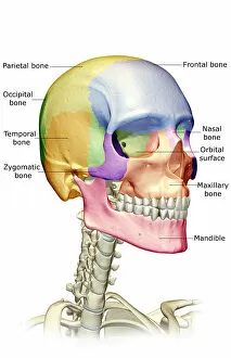
Background imageAnatomy Collection: anatomy, bone, bone structure, bone structure of the face, bone structure of the head

anatomy, bone, bone structure, bone structure of the face, bone structure of the head
anatomy, bone, bone structure, bone structure of the face, bone stru, Medical Image Collection, 87396684

Background imageAnatomy Collection: Anatomy of human knee joint

Anatomy of human knee joint

Background imageAnatomy Collection: Motor homunculus

Motor homunculus
This model shows what a mans body would look like if each part grew in proportion to the area of the cortex of the brain concerned with its movement

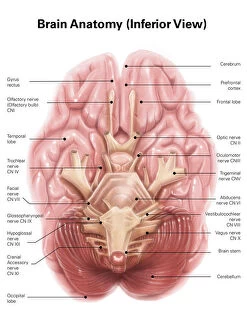
Background imageAnatomy Collection: Anatomy of human brain, inferior view

Anatomy of human brain, inferior view

Background imageAnatomy Collection: Facial muscles of the human face (with labels)

Facial muscles of the human face (with labels)

Background imageAnatomy Collection: Pen and ink studies, c1510, by Leonardo da Vinci of a human fetus

Pen and ink studies, c1510, by Leonardo da Vinci of a human fetus
LEONARDO: HUMAN FETUS. Pen and ink studies, c1510, by Leonardo da Vinci of a human fetus

Background imageAnatomy Collection: John Hunter, 1786. Artist: WO Geller

John Hunter, 1786. Artist: WO Geller
John Hunter, 1786. Scottish surgeon and anatomist whose series of lectures on the theory and practice of surgery attracted numerous students, including Edward Jenner, Astley Cooper

Background imageAnatomy Collection: Taste map of a human tongue, light green area sensitive to bitter taste

Taste map of a human tongue, light green area sensitive to bitter taste, yellow areas sensitive to sour taste, blue areas sensitive to salt taste, purple area sensitive to sweet taste

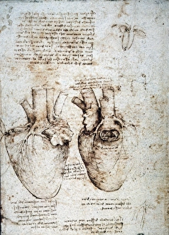
Background imageAnatomy Collection: Studies of an ox heart by Leonardo da Vinci, c1512, pen and ink

Studies of an ox heart by Leonardo da Vinci, c1512, pen and ink
LEONARDO: HEART, c1512. Studies of an ox heart by Leonardo da Vinci, c1512, pen and ink

Background imageAnatomy Collection: Giant octopus

Giant octopus
Plate 26 from Histoire naturelle des Mollusques by Count Georges Louis Leclerc de Buffon, 1805

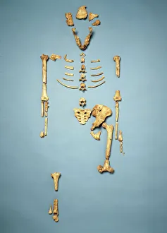
Background imageAnatomy Collection: Australopithecus afarensis (AL 288-1) (Lucy)

Australopithecus afarensis (AL 288-1) (Lucy)
A cast of the partial skeleton (nicknamed Lucy) of Australopithecus afarensis found at the Hadar, North East Ethiopia in 1974 by Donald Johanson

Background imageAnatomy Collection: The Anatomy Lesson of Dr Nicolaes Tulp, 1632. Artist: Rembrandt Harmensz van Rijn

The Anatomy Lesson of Dr Nicolaes Tulp, 1632. Artist: Rembrandt Harmensz van Rijn
The Anatomy Lesson of Dr Nicolaes Tulp, 1632. Rembrandt van Rhijn (1606-1669). Found in the collection of the The Mauritshuis, The Hague

Background imageAnatomy Collection: Human Heart Anatomy 1888

Human Heart Anatomy 1888
Illustrated Natural History of the Three Kingdoms Illustrierte Naturgeschichte der drei Reiche Franz StrAÔé¼ssles - Wilhelm Nitzschke, Stuttgart 1888

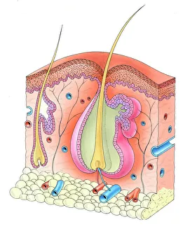
Background imageAnatomy Collection: Human hair follicle and skin

Human hair follicle and skin

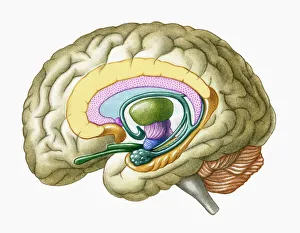
Background imageAnatomy Collection: Cross section illustration of human brain showing limbic system and primitive forebrain

Cross section illustration of human brain showing limbic system and primitive forebrain

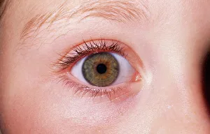
Background imageAnatomy Collection: Picture No. 10851598

Picture No. 10851598
Girlճ EYE - close-up of single hazel eye Date:

Background imageAnatomy Collection: Ara ararauna, blue-and-yellow macaw

Ara ararauna, blue-and-yellow macaw
Plate 41, a watercolour from the John Reeves Collection of Zoological Drawings from Canton, China

Background imageAnatomy Collection: Homo erectus (Sangiran 17), H. sapiens (?) H. neanderthalens

Homo erectus (Sangiran 17), H. sapiens (?) H. neanderthalens
L to R, cast of Homo erectus, (Sangiran 17), Sangiran, Java, 700, 000 years old; cast of recent Homo sapiens skull from South East Asia; cast of Neanderthal skull from La Ferrassie, France, 50

Background imageAnatomy Collection: Scanning Electron Micrograph (SEM): Tardigrade or ‘Water Bear Magnification x 1250 (A4 size: 29)

Scanning Electron Micrograph (SEM): Tardigrade or ‘Water Bear Magnification x 1250 (A4 size: 29. 7 cm width)
LRDS-129 Tardigrade or Water Bear Scanning Electron Micrograph (SEM) Ramazzotius oberhauseri Magnification x 1250 (A4 size: 29.7 cm width) Coloured by hand to enhance natural features

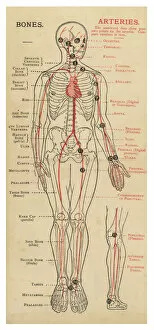
Background imageAnatomy Collection: Human body with bones and arteries

Human body with bones and arteries
A diagram of the human body, with details of bones and arteries

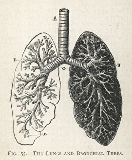
Background imageAnatomy Collection: Diagram of the lungs and bronchial tubes

Diagram of the lungs and bronchial tubes
A diagram of the lungs and bronchial tubes, with a section of the windpipe

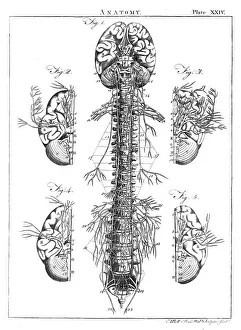
Background imageAnatomy Collection: Diagram of the human brain and spinal column

Diagram of the human brain and spinal column
A diagram of the brain and spinal column, including the vertebrae

Background imageAnatomy Collection: Front View of Lungs

Front View of Lungs
Detailed diagram of the lungs

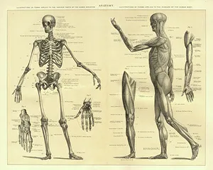
Background imageAnatomy Collection: Human Anatomy Skeleton and muscles of the body

Human Anatomy Skeleton and muscles of the body
Vintage engraving of Human Anatomy, the bones of the skeleton and muscles of the body, 1898

Background imageAnatomy Collection: Pharomachrus moccino, resplendent quetzal

Pharomachrus moccino, resplendent quetzal
Plate from John Goulds The Birds of New Guinea, (1875-1888). Hand coloured lithograph

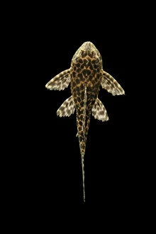
Background imageAnatomy Collection: Fish - Spotted Sailfin Pleco South America

Fish - Spotted Sailfin Pleco South America
BB-802 Fish - Spotted Sailfin Pleco South America Glyptoperichthys gibbiceps Brian Bevan Please note that prints are for personal display purposes only and may not be reproduced in any way

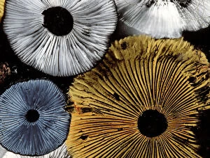
Background imageAnatomy Collection: Mushroom spores - close-up

Mushroom spores - close-up
LA-1754 Mushroom spores - close-up Jean Michel Labat Please note that prints are for personal display purposes only and may not be reproduced in any way

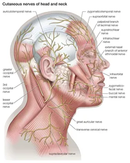
Background imageAnatomy Collection: Diagram of the cutaneous nerves of the head and neck

Diagram of the cutaneous nerves of the head and neck

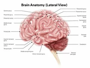
Background imageAnatomy Collection: Human brain anatomy, lateral view

Human brain anatomy, lateral view

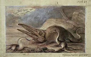
Background imageAnatomy Collection: Ichthyosaurus, Plesiosaurus

Ichthyosaurus, Plesiosaurus
Watercolour painting by Benjamin Waterhouse Hawkins, (1809-1889). These marine reptiles live in Europe duing the early Jurassic period about 200 millions years ago

Background imageAnatomy Collection: Anatomy / Skull / 1841

Anatomy / Skull / 1841
Illustration of the skull of a bushman of the Hottentot Tribe, Africa (side view)

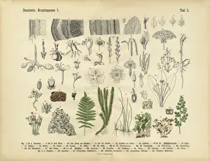
Background imageAnatomy Collection: Plant Anatomy, Victorian Botanical Illustration

Plant Anatomy, Victorian Botanical Illustration
Very Rare, Beautifully Illustrated Antique Engraved Victorian Botanical Illustration of Plant Anatomy: Plate 1, from Lehrbuch der praktischen Pflanzenkunde in Wort und Bild

Background imageAnatomy Collection: Pinus cembra, Arolla pine

Pinus cembra, Arolla pine - Coloured copper engraving by Warner based on a watercolour by Ferdinand Bauer. A.B. Lambert, A description of the genus Pinus, London, 1803, t. 23 Date: 1803

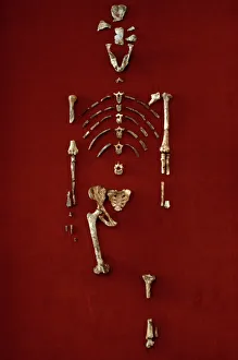
Background imageAnatomy Collection: Australopithecus afarensis (AL 288-1) (Lucy)

Australopithecus afarensis (AL 288-1) (Lucy)
A partial skeleton (nicknamed Lucy) of Australopithecus afarensis found at the Hadar, North East Ethiopia in 1974 by Donald Johanson

Background imageAnatomy Collection: Calyptorhynchus funereus, yellow-tailed black cockatoo

Calyptorhynchus funereus, yellow-tailed black cockatoo
Watercolour 137 by Thomas Watling from the Watling Collection titled Banksian Cockatoo, native name Kar ratt

Background imageAnatomy Collection: Ara macao, scarlet macaw

Ara macao, scarlet macaw
Plate 25, a watercolour from the John Reeves Collection of Zoological Drawings from Canton, China

Background imageAnatomy Collection: Ichthyosaurr, Plesiosaurus, Pterodactylus

Ichthyosaurr, Plesiosaurus, Pterodactylus
Duria Antiquior - A more ancient Dorset by Sir Henry Thomas De la Beche. This collection of marine and avian reptiles could be found in Europe during the early Jurassic period about 200 million years

Background imageAnatomy Collection: Xanthopan morganii praedicta, sphinx moth

Xanthopan morganii praedicta, sphinx moth
Darwins sphinx moth, found in Madagascar. The species as a whole is known as Morgans sphinx

Background imageAnatomy Collection: Ballyregan Bob, greyhound

Ballyregan Bob, greyhound
Photograph of Ballyregan Bob, the famous racing greyhound. Specimen on display at the Natural History Museum at Tring

Background imageAnatomy Collection: Calypte anna, Annas hummingbird

Calypte anna, Annas hummingbird

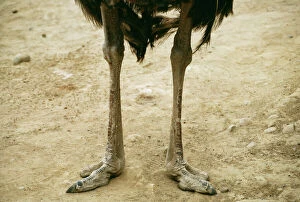
Background imageAnatomy Collection: Ostrich Legs & feet

Ostrich Legs & feet
KF-477 Ostrich - Legs & feet Strutio camelus Kenneth W Fink Please note that prints are for personal display purposes only and may not be reproduced in any way

Background imageAnatomy Collection: Great Hammerhead Shark - Swims towards the photographer. These sharks are considered dangerous

Great Hammerhead Shark - Swims towards the photographer. These sharks are considered dangerous. Coral Sea. Australia
VT-8479 Great Hammerhead SHARK - swimming towards the photographer Coral Sea. Australia Sphyrna mokarran These sharks are considered dangerous

Background imageAnatomy Collection: Greater Spotted Dogfish teeth and jaws. Teeth continuously replaced from behind

Greater Spotted Dogfish teeth and jaws. Teeth continuously replaced from behind. North sea and Mediterranean
PM-10075 Greater Spotted Dogfish - teeth and jaws, teeth continuously replaced from behind North sea and Mediterranean Scyliorhinus stellaris Pat Morris Please note that prints are for personal

Background imageAnatomy Collection: Gastric bypass procedure

Gastric bypass procedure
Detail, Anatomy, Diagram, Illustration, Medical, Stomach, Biology, Surgery, Weight Loss, Cut Out, White Background, Vertical, Artwork, Color Image, Close Up, Healthcare And Medicine, The Human Body

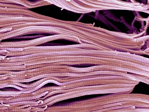
Background imageAnatomy Collection: Collagen, Scanning electron micrograph (SEM)

Collagen, Scanning electron micrograph (SEM)
Collagen. Scanning electron micrograph (SEM) of collagen bundles from the delicate connective tissue endoneurium. Endoneurium wraps around and between individual nerve fibres (axons)

Background imageAnatomy Collection: Scanning electron micrograph (SEM) of red blood cell

Scanning electron micrograph (SEM) of red blood cell
Anatomy, Biology, Blood Clot, Blood Vessel, Cell, Color Image, Fractur, SEM, 85758265

Background imageAnatomy Collection: Extinct marine reptiles

Extinct marine reptiles
Sheet 1 of a series of posters called Extinct Animals by Benjamin Waterhouse Hawkins c. 1862. This collection of marine reptiles lived during the Jurassic period between 200

Background imageAnatomy Collection: Diagram of inner ear showing auditory canal, eardrum, semicircular canals, cochlea, cochlea nerve

Diagram of inner ear showing auditory canal, eardrum, semicircular canals, cochlea, cochlea nerve, eustachian tube

Background imageAnatomy Collection: Illustration of human lymphatic system

Illustration of human lymphatic system

Background imageAnatomy Collection: Superior view of human brain with colored lobes and labels

Superior view of human brain with colored lobes and labels

Background imageAnatomy Collection: Nymphicus hollandicus, cockatiel

Nymphicus hollandicus, cockatiel
Plate 27 from Edward Lears Illustrations of the Family of Psittacidae or Parrots (1832). Hand coloured lithograph

Background imageAnatomy Collection: Cygnus atratus, black swan

Cygnus atratus, black swan
Watercolour 351 by the Port Jackson Painter from the Watling Collection

Background imageAnatomy Collection: Neophema pulchella, turquoise parrot

Neophema pulchella, turquoise parrot
Watercolour 132 by Thomas Watling from the Watling Collection titled Turcosine Parrot

Background imageAnatomy Collection: Phoeniconaias minor, lesser flamingo

Phoeniconaias minor, lesser flamingo
Pencil and watercolour drawing by Claude Gibney Finch-Davies (1918) from his African birds sketchbook

Background imageAnatomy Collection: Pelecanus erythrorynchos, American white pelican

Pelecanus erythrorynchos, American white pelican
Plate 311 from John James Audubons Birds of America, original double elephant folio (1835-38), hand-coloured aquatint. Engraved, printed and coloured by R. Havell (& Son), London

Background imageAnatomy Collection: Bubo coromandus, dusky eagle owl

Bubo coromandus, dusky eagle owl
A watercolour of the dusky eagle owl (Bubo coromandus) by an unknown artist, (c. 1840), part of the Lord Ashton Collection held in the Natural History Museum, London

Background imageAnatomy Collection: Medical / Anatomy / Blood

Medical / Anatomy / Blood
Circulation of the blood Date: Late 19th Century

Background imageAnatomy Collection: COMPARATIVE EMBRYOLOGY. Chart showing comparative embryology from a fish to a man

COMPARATIVE EMBRYOLOGY. Chart showing comparative embryology from a fish to a man, made by the Department of Comparative and Human Anatomy at the American Museum of Natural History, 1932

Background imageAnatomy Collection: Liver Fluke, microscope slide preparation of the whole animal

Liver Fluke, microscope slide preparation of the whole animal
PM-10632 Liver Fluke, microscope slide preparation of the whole animal. Fasciola hepatica Pat Morris Please note that prints are for personal display purposes only

Background imageAnatomy Collection: Scanning Electron Micrograph (SEM): Praying Mantis - Magnification x 30 (if print A4 size: 29)

Scanning Electron Micrograph (SEM): Praying Mantis - Magnification x 30 (if print A4 size: 29. 7 cm wide)
LRDS-392 Scanning Electron Micrograph (SEM): Praying Mantis Magnification x 30 (if print A4 size: 29.7 cm wide) Mantis religiosa David Spears (Last Refuge)

Background imageAnatomy Collection: Tiger Barb Fish

Tiger Barb Fish
LA-1325 Sumatra / Tiger BARB Fish Distribution: Sumatra and Borneo Puntius tetrazona Jean Michel Labat Please note that prints are for personal display purposes only

Background imageAnatomy Collection: Diagram of Ear / 1897

Diagram of Ear / 1897
EAR Diagram of the front view of the ear

Background imageAnatomy Collection: Normal knee, X-ray

Normal knee, X-ray
Normal knee. Coloured X-ray of the knee of a 44 year old woman

Background imageAnatomy Collection: Human circulatory system, hand-coloured engraving, published in 1861

Human circulatory system, hand-coloured engraving, published in 1861
The human circulatory system. Hand-coloured engraving, published in 1861

Background imageAnatomy Collection: Male and Female pelvis engraving 1896

Male and Female pelvis engraving 1896
Atlas d anatomie descriptive du corps humain C. Bonamy - Paul Broca Victor Masson et Fils Paris 1866

Background imageAnatomy Collection: Illustration of internal organs of a snake including heart, lung, intestines, pancreas

Illustration of internal organs of a snake including heart, lung, intestines, pancreas, kidney and testis

Background imageAnatomy Collection: Medical ilustration of an appendix with appendicitis

Medical ilustration of an appendix with appendicitis

Background imageAnatomy Collection: Cell types in the mammalian cerebellum: drawing, 1894, by the Spanish histologist Santiago Ramon y

Cell types in the mammalian cerebellum: drawing, 1894, by the Spanish histologist Santiago Ramon y Cajal (1852-1934)
NERVE CELLS, 1894. Cell types in the mammalian cerebellum: drawing, 1894, by the Spanish histologist Santiago Ramon y Cajal (1852-1934)


All Professionally Made to Order for Quick Shipping

"Unveiling the Intricacies of Anatomy: From Sensory Homunculus to Motor Homunculus" Step into the fascinating world of anatomy, where every detail tells a story. Explore the depths of human and animal structures as we delve into this captivating subject. Let's begin with Leonardo da Vinci's masterpiece, "Head of a Bear. " Created in 1480 but discovered in 1945, this artwork showcases da Vinci's unparalleled ability to capture anatomical accuracy. The intricate details reveal his keen observation skills and deep understanding of form. Moving on to our furry friends, the greyhound takes center stage. Discovering the anatomy of these majestic creatures unveils their remarkable speed and agility. From their sleek skeleton to their muscular build, it is evident why they are renowned for their racing abilities. But what about us humans? Enter the sensory homunculus – a visual representation that depicts how our brain perceives different body parts based on sensitivity levels. This map reveals intriguing insights into how our senses are distributed throughout our bodies. X-ray images provide another dimension to understanding anatomy. Take a glimpse at normal knees through an X-ray lens – marvel at the complexity hidden beneath our skin while appreciating its delicate balance between strength and flexibility. The backbone is often considered one of nature's most ingenious designs. Our human backbone includes ribs and pelvis, providing stability while allowing movement – truly an architectural marvel worth exploring further. Venturing deeper within ourselves, let us explore the intricacies of the human brain from an inferior view. Witness its complex network of connections responsible for controlling various bodily functions - truly awe-inspiring. As we journey back in time, we encounter hominid crania – remnants that shed light on our evolutionary history. These ancient skulls offer glimpses into early forms such as Australopithecus afarensis (AL 288-1), famously known as Lucy - bridging gaps between past and present.

© Copyright Media Storehouse, All Rights Reserved 2003 - 2024
Facebook     Pinterest     Card Payments     Reviews IO