
Sensory System Collection
The intricate and fascinating world of the sensory system is unveiled through these captivating images


All Professionally Made to Order for Quick Shipping
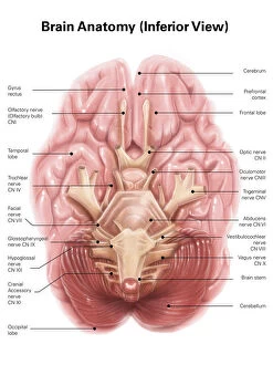
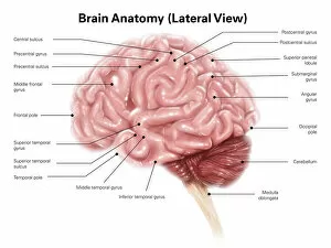
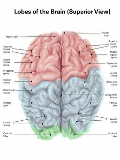
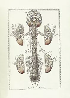
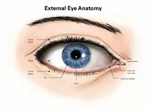
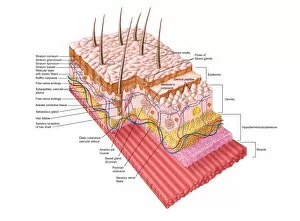
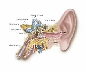
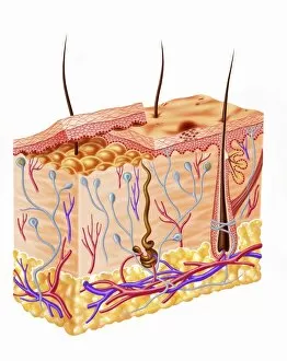
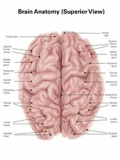
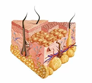
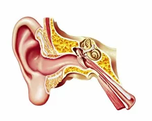
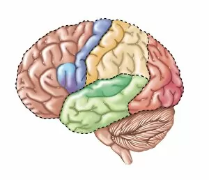
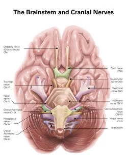
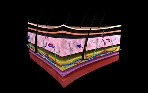
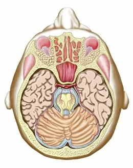
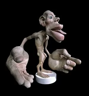
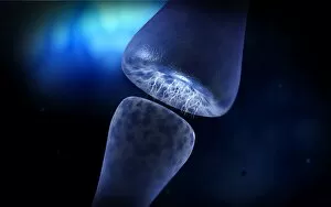
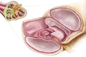
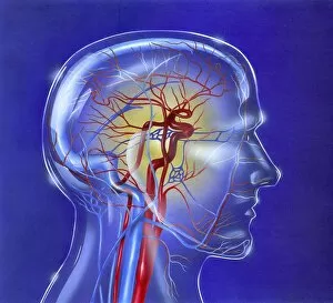
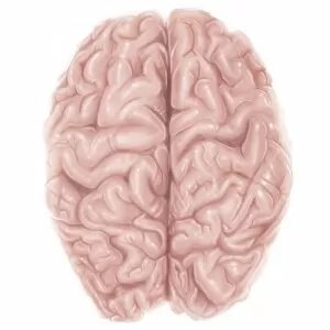
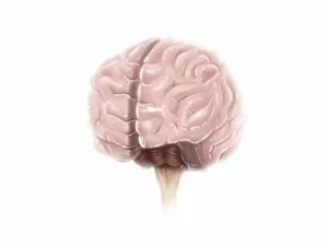
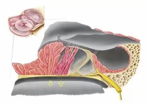
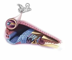
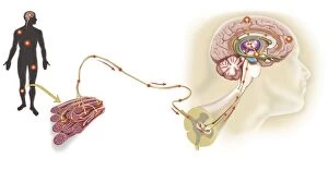
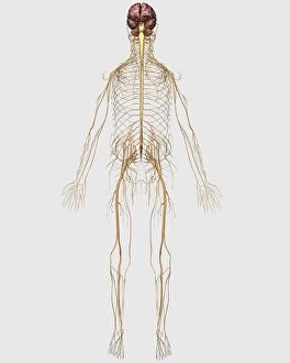
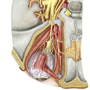
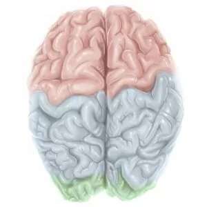
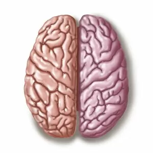
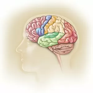
The intricate and fascinating world of the sensory system is unveiled through these captivating images. Starting with an inferior view of the anatomy of the human brain, we delve into the complex network that governs our senses. The motor and sensory homunculi reveal how different parts of our body are represented in our brain, showcasing the incredible precision with which our sensory experiences are processed. Moving to a superior view of the human brain, we encounter colored lobes and labels that highlight their distinct functions. This visual representation allows us to appreciate how each lobe contributes to our perception and interpretation of the world around us. As we explore further, Bartholomeo Eustachi's masterpiece "The Science of Human Anatomy" takes us on a journey through various anatomical structures. From external anatomy of the human eye with detailed labels, we gain insight into its remarkable ability to capture light and form images. Next, we unravel the secrets hidden beneath our skin as we examine the anatomy of this vital organ. Its complexity becomes evident as layers upon layers work harmoniously to protect us while allowing for tactile sensations. Venturing deeper into auditory perception, we encounter both external auditory canal and cochlear duct in exquisite detail. These illustrations shed light on how sound waves travel through these pathways before reaching our brains – enabling us to hear melodies or whispers alike. Anatomical pathways leading to innervation in lacrimal gland offer insights into another aspect: tears - not only expressions but also essential for maintaining ocular health. Finally, a superior view reveals brain surface anatomy adorned with informative labels. It showcases key regions responsible for cognition, memory formation, emotion regulation – all crucial components influencing how we perceive stimuli from within ourselves or from outside sources. These captivating visuals provide glimpses into one's understanding about sensory systems' inner workings—reminding us just how marvelous it is that such intricate processes occur seamlessly within each one of us every day.