
Aural Collection
"Aural: Exploring the Symphony of Sound Through Time and Art" Step back in time to the early days of communication


All Professionally Made to Order for Quick Shipping
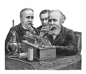
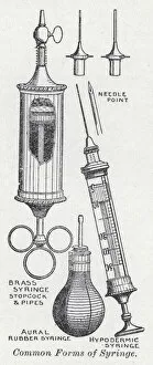
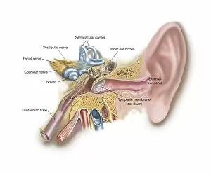
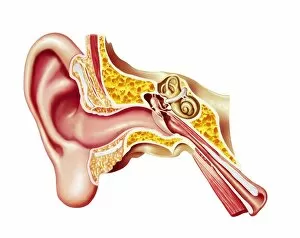
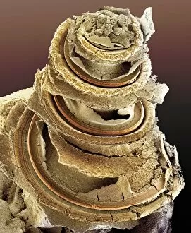
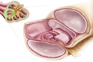
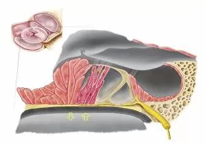
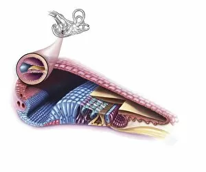
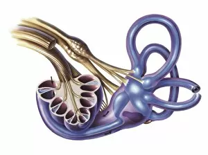
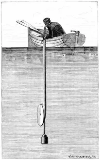
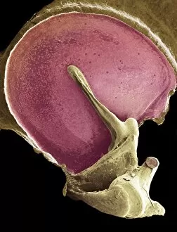
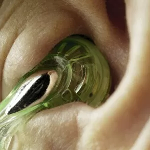
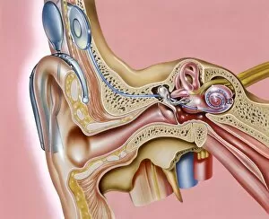
"Aural: Exploring the Symphony of Sound Through Time and Art" Step back in time to the early days of communication, where the telephone was a marvel that connected distant voices. Uncover historical artwork that captures the essence experiences, from mesmerizing landscapes to intimate portraits. Witness a doctor delicately syringing a patient's ear, providing relief and restoring harmony to their auditory world. Discover common forms of syringes depicted in lithographs, showcasing the evolution of medical practices for ear care. Immerse yourself in "The Ingle Nook, " an enchanting engraving that invites you to cozy up by the fire and indulge in captivating stories told through whispers and melodies. In the evening, as she lies on her bed, she rereads the heartfelt letter from her artilleryman at war - every word resonating deep within her soul like a symphony composed just for her. Delve into the intricate anatomy of our cochlear ducts, unraveling how these delicate structures transform vibrations into beautiful melodies we perceive as sound. Explore the external auditory canal with labeled diagrams, unlocking secrets hidden within this gateway to our sonic universe. Journey further into our inner ear and sinuses' complex anatomy – marvel at its intricacies designed to capture every note and rhythm life has to offer. Peek inside a cutaway diagram revealing layers upon layers of mechanisms working harmoniously together within our human ears – nature's masterpiece finely tuned for auditory perfection. Behold an astonishing SEM image capturing the Organ of Corti – witness its microscopic beauty responsible for translating sound waves into electrical signals that dance along our neural pathways like musical notes on sheet music.