Fraud Blocker Skip to main content

Magnification Collection

Magnification: Revealing the Hidden World From the intricate patterns of a snowflake to the delicate structure of a human cell

Background imageMagnification Collection: Mandelbrot fractal

Mandelbrot fractal. Computer-generated image derived form a Mandelbrot Set

Background imageMagnification Collection: Crysotile asbestos

Crysotile asbestos
Scanning electron micrograph of 5-Fold symmetry in crysotile asbestos. Magnification on the 5 x4 transparency = X 600, 000

Background imageMagnification Collection: Medical ilustration of an appendix with appendicitis

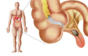
Medical ilustration of an appendix with appendicitis

Background imageMagnification Collection: Liver

Liver
Scanning electron microscope (SEM) image of a section through a liver (x 7000), an organ that has over 500 functions in the human body (x 800)

Background imageMagnification Collection: Human cell

Human cell
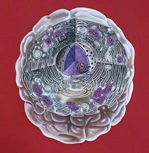
Illustration of a highly magnified section through a human cell. Page 8 from Human Biology, 1977

Background imageMagnification Collection: Julia fractal

Julia fractal. Computer-generated image derived form a Julia Set

Background imageMagnification Collection: Taraxacum officinale, dandelion (fruiting head)

Taraxacum officinale, dandelion (fruiting head)
Scanning electron microscope image showing a vertical section through an unripe fruiting head of a dandelion in the yellow flower stage. Colour added artificially by computer

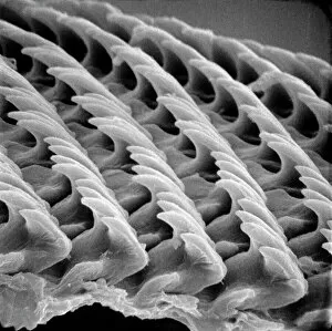
Background imageMagnification Collection: Snail teeth

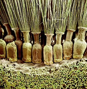
Snail teeth

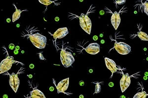
Background imageMagnification Collection: Water fleas (Daphnia sp. ) and a green algae (Volvox aureus) in water from a garden pond

Water fleas (Daphnia sp. ) and a green algae (Volvox aureus) in water from a garden pond
Water fleas (Daphnia sp.) and a green algae (Volvox aureus) in water from a garden pond. Derbyshire, UK. September. Digital composite

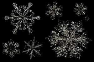
Background imageMagnification Collection: Different Snowflakes showing range in size and pattern, magnified under microscope

Different Snowflakes showing range in size and pattern, magnified under microscope, from Lilehammer, Norway. Digital composite

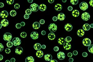
Background imageMagnification Collection: Chlorophyte or green alga (Volvox aureus) in pond water. UK. The larger spheres are approximately 0

Chlorophyte or green alga (Volvox aureus) in pond water. UK. The larger spheres are approximately 0.5mm in diameter. Digital composite image

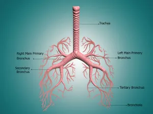
Background imageMagnification Collection: Anatomy of the bronchus and bronchial tubes

Anatomy of the bronchus and bronchial tubes
Digital illustration showing the structure of the bronchus and bronchial tubes. These pathways carry air into the lungs and carbon dioxide out
Background imageMagnification Collection: Circuit board, artwork

Circuit board, artwork
Circuit board. Computer artwork depicting city scape made of electronic circuits

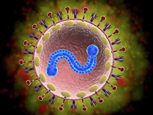
Background imageMagnification Collection: Microscopic view of human respiratory syncytial virus

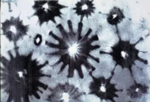
Microscopic view of human respiratory syncytial virus (RSV). RSV causes respiratory tract infection of the lungs and breathing passages

Background imageMagnification Collection: Sugar crystals, ordinary table sugar, photomicrography

Sugar crystals, ordinary table sugar, photomicrography

Background imageMagnification Collection: Human fetus inside amniotic sac

Human fetus inside amniotic sac

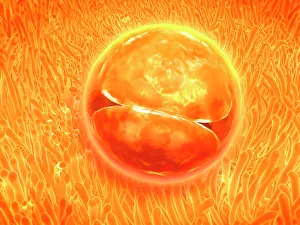
Background imageMagnification Collection: Embryo development 24-36 hours after fertilization

Embryo development 24-36 hours after fertilization

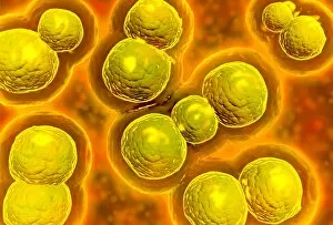
Background imageMagnification Collection: Microscopic view of chlamydia

Microscopic view of chlamydia. Chlamydia is a common sexually transmitted disease (STD) caused by a bacterium

Background imageMagnification Collection: Scyliorhinus canicula, dogfish

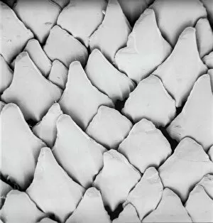
Scyliorhinus canicula, dogfish
Scanning electron microscope (SEM) image of the scales of a dogfish (x 40)

Background imageMagnification Collection: False-coloured SEM (Scanning Electron Micrograph) of a Tardigrade (Tardigrada) amongst moss

False-coloured SEM (Scanning Electron Micrograph) of a Tardigrade (Tardigrada) amongst moss, from Peak District National Park, Derbyshire, UK. This tardigrade is approximately 0.1 millimetres long

Background imageMagnification Collection: T2 cell culture

T2 cell culture
Scanning electron microscope image showing a T2 cell culture (x 4K)

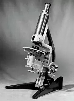
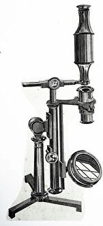
Background imageMagnification Collection: Rosenhain optical microscope

Rosenhain optical microscope. The viewing lens (eyepiece, upper right), focusing wheel (lower right) and objective lens (centre) are seen

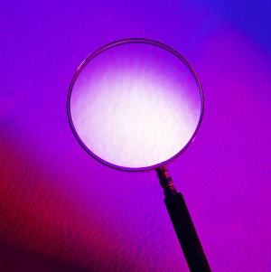
Background imageMagnification Collection: Magnifying glass

Magnifying glass. The glass lens is polished to an exact shape to produce a clear image. It magnifies the view of an object because the shape of the lens is convex
Background imageMagnification Collection: Fractal cloud, computer artwork

Fractal cloud, computer artwork
Fractal cloud. Computer graphic image of a cloud formed by fractal forms. This has been coloured using mathematical operations similar to those found in fractal geometry

Background imageMagnification Collection: Light Micrograph (LM) of a longitudinal section of stem showing xylem elements of Crown of Thorns

Light Micrograph (LM) of a longitudinal section of stem showing xylem elements of Crown of Thorns (Euphorbia splendens), magnification x1200

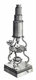
Background imageMagnification Collection: Microscope by Adams of London

Microscope by Adams of London
5311766 Microscope by Adams of London; (add.info.: Illustration of a microscope by Adams of London. Dated circa 1788.); Universal History Archive/UIG.

Background imageMagnification Collection: Three pillar microscope

Three pillar microscope
5311760 Three pillar microscope; (add.info.: Illustration of a 'Three pillar microscope' of the type produced by Edmund Culpeper (1660-1738) circa 1730.); Universal History Archive/UIG.

Background imageMagnification Collection: Compound microscope

Compound microscope
5311762 Compound microscope; (add.info.: Illustration depicting a compound microscope by Adams of London. Dated circa 1788.); Universal History Archive/UIG.

Background imageMagnification Collection: Focus stacked, inverted image of Diatoms on a microscope slide

Focus stacked, inverted image of Diatoms on a microscope slide. Diatoms are single-celled algae which produce approximately 25% of the oxygen we breathe as well as being responsible for approximately

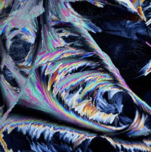
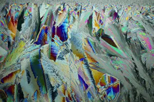
Background imageMagnification Collection: Sheet ice crystals viewed by polarised light

Sheet ice crystals viewed by polarised light

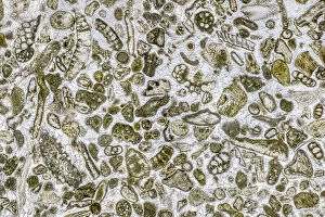
Background imageMagnification Collection: Limestone microfossils in a 0. 03mm-thick slice of fossil-rich limestone viewed at high

Limestone microfossils in a 0. 03mm-thick slice of fossil-rich limestone viewed at high magnification
Limestone microfossils in a 0.03mm-thick slice of fossil-rich limestone viewed at high magnification, image area is 3mm across in real life

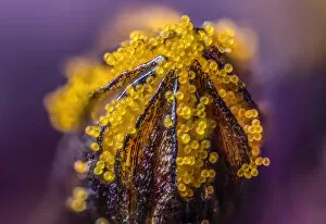
Background imageMagnification Collection: African daisy (Osteospermum jucundum) at approx 10x magnification

African daisy (Osteospermum jucundum) at approx 10x magnification, composite flower showing individual florets full of golden pollen grains, Monmouthshire, Wales, UK, July

Background imageMagnification Collection: Plankton at night, Palau, Philippine Sea

Plankton at night, Palau, Philippine Sea

Background imageMagnification Collection: Ice crystals viewed by polarised light

Ice crystals viewed by polarised light

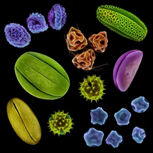
Background imageMagnification Collection: Various pollen grains from several species of UK plant. False coloured scanning electron micrograph

Various pollen grains from several species of UK plant. False coloured scanning electron micrograph. Digital composite

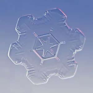
Background imageMagnification Collection: Snowflake magnified under microscope, Lilehammer, Norway

Snowflake magnified under microscope, Lilehammer, Norway

Background imageMagnification Collection: Northern Jungle Queen (Stichophthalma camadeva) female, India

Northern Jungle Queen (Stichophthalma camadeva) female, India

Background imageMagnification Collection: Red campion (Silene dioica) seeds, microscopic view

Red campion (Silene dioica) seeds, microscopic view

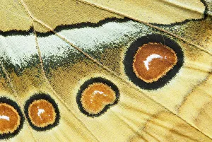
Background imageMagnification Collection: Abstract close-up of butterfly wing scales

Abstract close-up of butterfly wing scales

Background imageMagnification Collection: Macro dandelion seed

Macro dandelion seed

Background imageMagnification Collection: False-coloured scanning electron micrograph of a Thrip's (Thysanoptera) head

False-coloured scanning electron micrograph of a Thrip's (Thysanoptera) head, the bulging compound eyes can be seen on either side of the head

Background imageMagnification Collection: Soil bacteria cultured on nutrient agar from a soil sample collected in Via Gellia Woods, Derbyshire

Soil bacteria cultured on nutrient agar from a soil sample collected in Via Gellia Woods, Derbyshire. Image size in real life 10mm across. UK. March

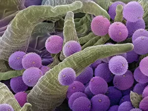
Background imageMagnification Collection: Pollen grains on the style of a Harebell (Campanula rotundifolia), Hoe Grange Nature Reserve

Pollen grains on the style of a Harebell (Campanula rotundifolia), Hoe Grange Nature Reserve, Derbyshire, UK. August

Background imageMagnification Collection: Harebell (Campanula rotundifolia) pollen grain, false-coloured Scanning Electron Micrograph

Harebell (Campanula rotundifolia) pollen grain, false-coloured Scanning Electron Micrograph

Background imageMagnification Collection: Projector for magnifying microscopic telegraph messages (litho)

Projector for magnifying microscopic telegraph messages (litho)
5997198 Projector for magnifying microscopic telegraph messages (litho) by European School, (19th century); Private Collection; (add.info.: Projector for magnifying microscopic telegraph messages)

Background imageMagnification Collection: Magic Glasses, 1891 (oil on canvas)

Magic Glasses, 1891 (oil on canvas)
2652545 Magic Glasses, 1891 (oil on canvas) by Elmer, Edwin Romanzo (1850-1923); 35.6x25.4 cm; Shelburne Museum, Vermont, USA; eShelburne Museum; Museum Purchase; American, out of copyright

Background imageMagnification Collection: Chest Microscope, with selection of ivory sliders and list, c. 1800 (mixed media)

Chest Microscope, with selection of ivory sliders and list, c. 1800 (mixed media)
MFR348314 Chest Microscope, with selection of ivory sliders and list, c.1800 (mixed media) by English School, (18th-19th century); Private Collection; English, out of copyright

Background imageMagnification Collection: Microscope (wood, metal & glass) (see also 1113963)

Microscope (wood, metal & glass) (see also 1113963)
1113962 Microscope (wood, metal & glass) (see also 1113963) by French School; Private Collection; (add.info.: cased by Le Canu a Rouen); Photo eChristies Images; French, out of copyright


All Professionally Made to Order for Quick Shipping

Magnification: Revealing the Hidden World From the intricate patterns of a snowflake to the delicate structure of a human cell, magnification allows us to explore and unravel the mysteries of our world. Peering through the lens, we uncover secrets that would otherwise remain unseen. Take for instance Crysotile asbestos, its fibrous nature exposed under intense magnification, reminding us of its hidden dangers lurking within. In medical illustrations, an appendix with appendicitis comes alive as we zoom in closer. The inflamed tissues become vividly apparent, highlighting the urgency for surgical intervention. Moving deeper into our bodies, we discover the liver - a complex organ responsible for countless vital functions. Under magnification, its intricate network of cells and blood vessels reveals itself like an elaborate cityscape bustling with activity. But it's not just within ourselves that magnification unveils wonders; it extends beyond to nature's realm. A dandelion's fruiting head transforms into a mesmerizing display when observed up close – each tiny seed ready to embark on its own journey through wind and time. Fractals captivate our imagination as well – from Julia fractals with their infinite complexity to Mandelbrot fractals' self-repeating patterns. These mathematical marvels remind us of nature's innate ability to create beauty even in abstract forms. Anton van Leeuwenhoek revolutionized science by observing animalcules through his microscope in 1795. His discoveries opened up new frontiers in microbiology and forever changed our understanding of life at microscopic levels. Even seemingly ordinary creatures like snails hold surprises when examined closely; their teeth reveal intricate structures designed for efficient feeding and survival. Ponds teem with life too - water fleas gracefully glide alongside green algae like Volvox aureus. Magnified images transport us into this miniature ecosystem where every organism plays a crucial role in maintaining balance and harmony. And finally, we come full circle with snowflakes.