
Diencephalon Collection
The diencephalon, a vital part of the human brain anatomy, is often overlooked but plays a crucial role in various functions


All Professionally Made to Order for Quick Shipping
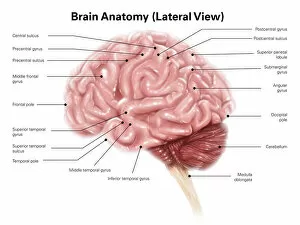
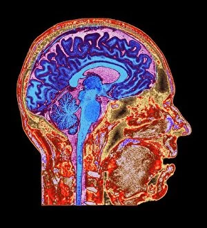
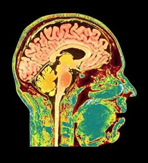
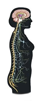
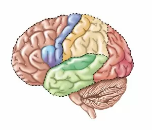
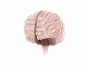
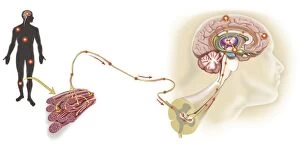
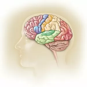
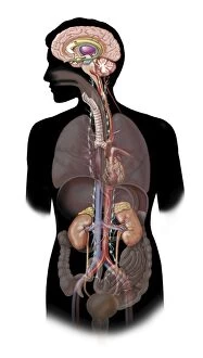
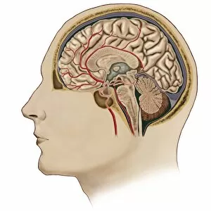
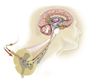
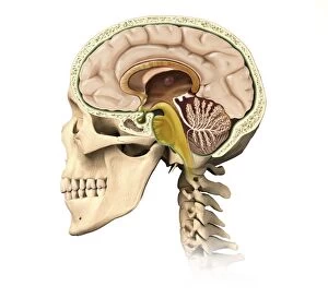
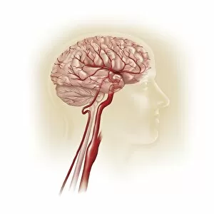
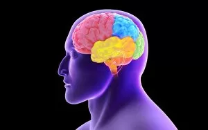
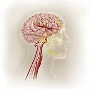
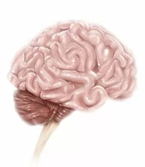
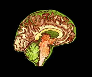
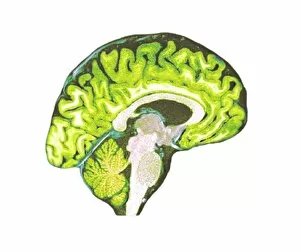
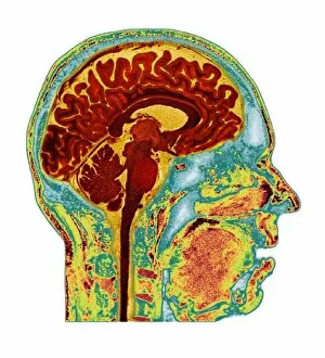
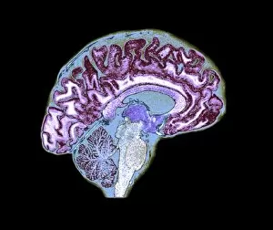
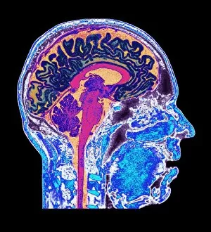
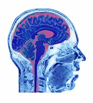
The diencephalon, a vital part of the human brain anatomy, is often overlooked but plays a crucial role in various functions. When viewing the lateral view of a normal human brain through MRI scans such as C016 / 8845 and C016 / 8850, one can observe the intricate structures within this region. In artwork depicting head and neck anatomy, we can see how the diencephalon fits into the larger context of our body. MRI scan C016 / 8843 provides further insight into the normal human brain's complexity, showcasing its detailed surface anatomy. The diencephalon is not only responsible for autonomic nervous system regulation but also contributes to our limbic system, which controls emotions and memory formation. By examining brain surface anatomy with labeled regions or observing side views highlighting functional lobes, we gain a deeper understanding of how different parts interact within this remarkable organ. Even during foetal brain development depicted through artwork, it becomes evident that the diencephalon undergoes significant changes to support future cognitive abilities. Furthermore, an injured muscle sends pain messages via sensory nerves along pathways that involve the diencephalon. This highlights its involvement in processing and interpreting sensations throughout our bodies. Lastly, let us not forget about another important structure related to the diencephalon -the pituitary gland- whose anatomical details are worth exploring.