Fraud Blocker Skip to main content

Telencephalon Collection

The telencephalon, also known as the cerebral hemispheres, is a fascinating part of the human brain

Background imageTelencephalon Collection: Human brain anatomy, lateral view

Human brain anatomy, lateral view

Background imageTelencephalon Collection: Normal human brain, MRI scan C016 / 8845

Normal human brain, MRI scan C016 / 8845
Brain. Coloured magnetic resonance imaging (MRI) scan of a sagittal section through a patients head showing a healthy human brain

Background imageTelencephalon Collection: Normal human brain, MRI scan C016 / 8850

Normal human brain, MRI scan C016 / 8850
Brain. Coloured magnetic resonance imaging (MRI) scan of a sagittal section through a patients head showing a healthy human brain

Background imageTelencephalon Collection: Normal human brain, MRI scan C016 / 8843

Normal human brain, MRI scan C016 / 8843
Brain. Coloured magnetic resonance imaging (MRI) scan of a sagittal section through a healthy human brain. The highly folded area (brown) is the cerebrum, which is responsible for conscious thought

Background imageTelencephalon Collection: Human body showing autonomic nervous system and limbic system

Human body showing autonomic nervous system and limbic system
Side view of human body showing autonomic nervous system and limbic system within the brain. Green are parasympathetic nerves. Blue are sympathetic nerves

Background imageTelencephalon Collection: Brain surface anatomy, with labels

Brain surface anatomy, with labels

Background imageTelencephalon Collection: Side view of the human brain showing the functional lobes

Side view of the human brain showing the functional lobes

Background imageTelencephalon Collection: Foetal brain development, artwork

Foetal brain development, artwork. During the 4th week (upper left) the neural tube begins to differentiate into a spinal cord (green), forebrain (blue), midbrain (grey) and hindbrain (orange)

Background imageTelencephalon Collection: Digital illustration of Electric Ray showing electricity and magnetism in cerebellum, lobus electric

Digital illustration of Electric Ray showing electricity and magnetism in cerebellum, lobus electric

Background imageTelencephalon Collection: Brain surface anatomy

Brain surface anatomy

Background imageTelencephalon Collection: Pathway of a pain message via sensory nerve in injured muscle

Pathway of a pain message via sensory nerve in injured muscle, to pain gate in spinal cord to limbic system, frontal cortex and sensory cortex in the human brain

Background imageTelencephalon Collection: Anatomy of pituitary gland

Anatomy of pituitary gland

Background imageTelencephalon Collection: Representation of how our senses affect our thoughts

Representation of how our senses affect our thoughts

Background imageTelencephalon Collection: Side view of the human head showing the functional lobes of the brain

Side view of the human head showing the functional lobes of the brain
Side view of the human head showing the functional lobes of the human brain

Background imageTelencephalon Collection: The sympathetic nervous system and the organs of fight-or-flight response

The sympathetic nervous system and the organs of fight-or-flight response

Background imageTelencephalon Collection: View of limbic system as seen from directly above the head

View of limbic system as seen from directly above the head

Background imageTelencephalon Collection: Cross section of brain with arteries

Cross section of brain with arteries

Background imageTelencephalon Collection: Human brain showing the layout of the sensory cortex

Human brain showing the layout of the sensory cortex
Coronal section through human brain showing the layout of the sensory cortex

Background imageTelencephalon Collection: Endorphins released in the hypothalamus travel down the spinal cord

Endorphins released in the hypothalamus travel down the spinal cord
Serotonin released in the brain travels down the spinal cord to close the pain gates and block pain messages

Background imageTelencephalon Collection: Sagittal view of human brain showing the corpus callosum

Sagittal view of human brain showing the corpus callosum

Background imageTelencephalon Collection: Cutaway view of human skull showing brain details, side view

Cutaway view of human skull showing brain details, side view

Background imageTelencephalon Collection: Side view of human brain showing internal carotid artery

Side view of human brain showing internal carotid artery

Background imageTelencephalon Collection: X-ray view of human face, profile view

X-ray view of human face, profile view

Background imageTelencephalon Collection: Detail of ateries of the human head and the trigeminal nerve

Detail of ateries of the human head and the trigeminal nerve

Background imageTelencephalon Collection: Human brain anatomy, lateral view, with labels

Human brain anatomy, lateral view, with labels

Background imageTelencephalon Collection: Normal human brain, MRI scan C016 / 8840

Normal human brain, MRI scan C016 / 8840
Brain. Coloured magnetic resonance imaging (MRI) scan of a sagittal section through a healthy human brain. The highly folded area (yellow) is the cerebrum, which is responsible for conscious thought

Background imageTelencephalon Collection: Normal human brain, MRI scan C016 / 8848

Normal human brain, MRI scan C016 / 8848
Brain. Coloured magnetic resonance imaging (MRI) scan of a sagittal section through a patients head showing a healthy human brain

Background imageTelencephalon Collection: Normal human brain, MRI scan C016 / 8837

Normal human brain, MRI scan C016 / 8837
Brain. Coloured magnetic resonance imaging (MRI) scan of a sagittal section through a healthy human brain. The highly folded area (orange) is the cerebrum, which is responsible for conscious thought

Background imageTelencephalon Collection: Normal human brain, MRI scan C016 / 8841

Normal human brain, MRI scan C016 / 8841
Brain. Coloured magnetic resonance imaging (MRI) scan of a sagittal section through a healthy human brain. The highly folded area (red and yellow) is the cerebrum

Background imageTelencephalon Collection: Normal human brain, MRI scan C016 / 8835

Normal human brain, MRI scan C016 / 8835
Brain. Coloured magnetic resonance imaging (MRI) scan of a sagittal section through a healthy human brain. The highly folded area across top is the cerebrum

Background imageTelencephalon Collection: Normal human brain, MRI scan C016 / 8846

Normal human brain, MRI scan C016 / 8846
Brain. Coloured magnetic resonance imaging (MRI) scan of a sagittal section through a patients head showing a healthy human brain

Background imageTelencephalon Collection: Normal human brain, MRI scan C016 / 8834

Normal human brain, MRI scan C016 / 8834
Brain. Coloured magnetic resonance imaging (MRI) scan of a sagittal section through a healthy human brain. The highly folded area (orange) is the cerebrum, which is responsible for conscious thought

Background imageTelencephalon Collection: Normal human brain, MRI scan C016 / 8849

Normal human brain, MRI scan C016 / 8849
Brain. Coloured magnetic resonance imaging (MRI) scan of a sagittal section through a patients head showing a healthy human brain

Background imageTelencephalon Collection: Normal human brain, MRI scan C016 / 8836

Normal human brain, MRI scan C016 / 8836
Brain. Coloured magnetic resonance imaging (MRI) scan of a sagittal section through a healthy human brain. The highly folded area across top is the cerebrum

Background imageTelencephalon Collection: Normal human brain, MRI scan C016 / 8844

Normal human brain, MRI scan C016 / 8844
Brain. Coloured magnetic resonance imaging (MRI) scan of a sagittal section through a patients head showing a healthy human brain

Background imageTelencephalon Collection: Normal human brain, MRI scan C016 / 8839

Normal human brain, MRI scan C016 / 8839
Brain. Coloured magnetic resonance imaging (MRI) scan of a sagittal section through a healthy human brain. The highly folded area across top is the cerebrum

Background imageTelencephalon Collection: Normal human brain, MRI scan C016 / 8847

Normal human brain, MRI scan C016 / 8847
Brain. Coloured magnetic resonance imaging (MRI) scan of a sagittal section through a patients head showing a healthy human brain

Background imageTelencephalon Collection: Cerebrum, light micrograph C014 / 1399

Cerebrum, light micrograph C014 / 1399
Cerebrum. Light micrograph of a section through tissue from the cerebrum, showing neurons (nerve cells, purple). The cerebrum is the largest part of the brain

Background imageTelencephalon Collection: Cerebrum, light micrograph C014 / 1400

Cerebrum, light micrograph C014 / 1400
Cerebrum. Light micrograph of a section through tissue from the cerebrum, showing neurons (nerve cells, purple). The cerebrum is the largest part of the brain

Background imageTelencephalon Collection: Cerebrum, light micrograph C014 / 0636

Cerebrum, light micrograph C014 / 0636
Cerebrum. Light micrograph of a section through part of the cerebrum of the brain. Seen here are the three main layers. The outer grey matter comprises the molecular layer (light pink)

Background imageTelencephalon Collection: Brain, artwork C014 / 6803

Brain, artwork C014 / 6803
Brain, computer artwork. At bottom is the brainstem, which controls automatic functions, sleep and arousal and relays messages from the brain to the spinal cord

Background imageTelencephalon Collection: Central nervous system, artwork

Central nervous system, artwork
Central nervous system. Computer artwork showing the brain, cranial nerves and the top portion of the spinal cord. At centre is the pituitary gland (small, spherical), which secretes hormones

Background imageTelencephalon Collection: Neural stem cells

Neural stem cells. Light micrograph of self- renewing neural stem cells taken from the brain of a mouse embryo. These stem cells can differentiate into neurons (nerve cells)

Background imageTelencephalon Collection: Frontal lobe in the brain, artwork

Frontal lobe in the brain, artwork
Frontal lobe in the brain. Computer artwork of a persons head showing the left hemisphere of the brain inside. The highlighted area shows the left frontal lobe

Background imageTelencephalon Collection: Frontal lobes in the brain, artwork

Frontal lobes in the brain, artwork
Frontal lobes in the brain. Computer artwork of a persons head showing the brain inside. The highlighted area shows the frontal lobes

Background imageTelencephalon Collection: Human brain, artwork

Human brain, artwork
Human brain anatomy. Computer artwork of a persons head showing the brain with the right hemisphere removed. The following regions can be seen: outer cortex (red) somatosensory cortex (lighter)

Background imageTelencephalon Collection: Cingulate gyrus in the brain, artwork

Cingulate gyrus in the brain, artwork
Temporal lobe in the brain. Computer artwork of a persons head showing the left hemisphere of the brain inside. The highlighted area shows the left temporal lobe


All Professionally Made to Order for Quick Shipping

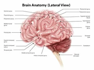
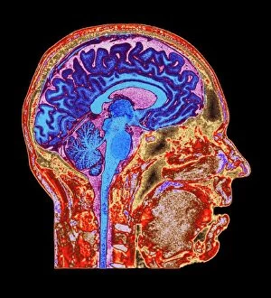
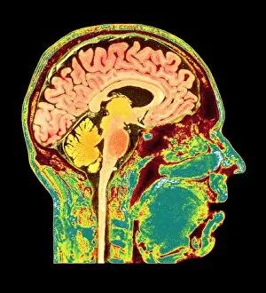
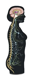
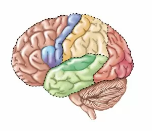
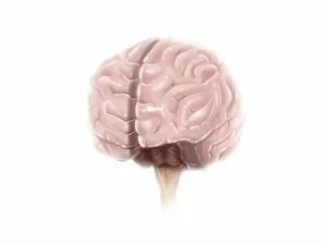
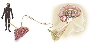
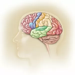
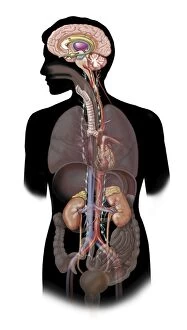
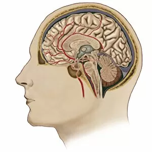
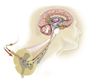
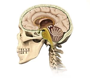
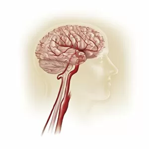
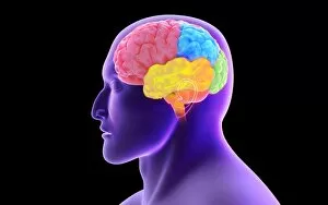
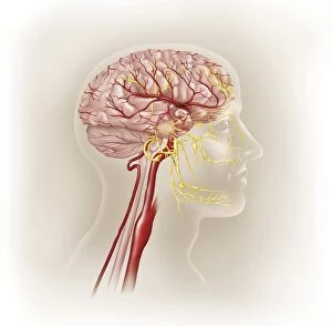
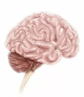
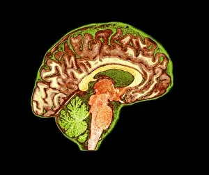
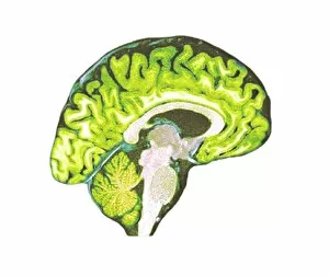
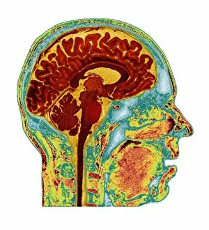
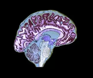
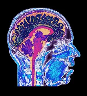
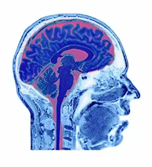
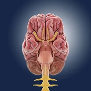
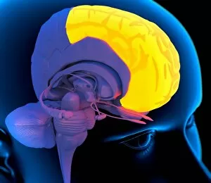
The telencephalon, also known as the cerebral hemispheres, is a fascinating part of the human brain. In this captivating image, we are presented with a lateral view of the telencephalon in all its intricate glory. The MRI scans C016/8845, C016/8850, and C016/8843 provide us with detailed visuals of a normal human brain. As we delve deeper into understanding this complex organ, we discover that it plays an integral role in our autonomic nervous system and limbic system. This connection between our body's involuntary functions and emotions highlights the significance of the telencephalon in regulating our responses to various stimuli. Examining the surface anatomy of the brain reveals labeled structures that aid in comprehending its functionality. Additionally, observing a side view allows us to identify different lobes responsible for specific cognitive processes. Intriguingly, even during fetal development, the telencephalon undergoes remarkable changes. Artwork depicting foetal brain development showcases how this region evolves over time. Expanding beyond humans' realm alone, an electric ray provides insight into electricity and magnetism within another area called lobus electric found within cerebellum through digital illustration. Furthermore, exploring pain pathways via sensory nerves sheds light on how messages travel from injured muscles to reach our brains—highlighting yet another aspect influenced by the telencephalon. Lastly but not least important is understanding pituitary gland anatomy since it works closely with other parts of the brain including Telencephalon which controls many vital bodily functions regulated by hormones produced by Pituitary Gland Overall these images offer glimpses into both macroscopic and microscopic aspects related to Telencephalon's structure and function—a testament to its crucial role in shaping our cognition and overall well-being