Poster Print > Science > SEM
Poster Print : Skeletal muscle fibre


![]()
Poster Prints from Science Photo Library
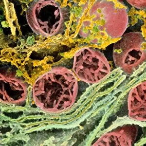
Skeletal muscle fibre
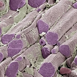
Skeletal muscle fibre. Coloured scanning electron micrograph (SEM) of skeletal muscle fibre. This type of muscle is striated. Muscle cells contain cylindrical organelles in the form of bundles of filaments (orange, myofibrils). Each filament can be thick (made up of myosin) or thin (made up of actin). In the case of skeletal (and cardiac) muscle, the filaments have a specific length, less than the length of the muscle cell. They are organised into repeating subunits (sarcomeres) along the length of the muscle cell. The resulting myofibrils run parallel to each other, causing the cell to appear striped (striated). The muscle is surrounded by connective tissue (loose strands)
Science Photo Library features Science and Medical images including photos and illustrations
Media ID 6448559
© SUSUMU NISHINAGA/SCIENCE PHOTO LIBRARY
Actin Connective Tissue Endomysium False Colour Fibre Fibres Filament Filaments Muscles Myofibril Myofibrils Myosin Orange Repeating Sarcomere Sarcomeres Skeletal Strand Strands Striated Striped Sub Unit Subunits Thick Thin Tissue False Coloured Repeat
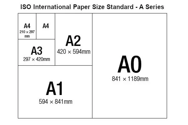
A2 (59.4 x 42cm) Poster Print
Discover the intricacy of life with our Media Storehouse Skeletal Muscle Fibre Poster Print. This captivating image, sourced from Science Photo Library, showcases a stunning coloured scanning electron micrograph (SEM) of a skeletal muscle fibre. Witness the striation and complex structure of this essential component of the human body. Bring the wonders of science into your workspace or home decor with our high-quality, vibrant poster prints. Ideal for educators, students, or anyone with an appreciation for the beauty of the natural world.
A2 Poster (59.4 x 42cm, 23.4" x 16.5" inches) printed on 170gsm Satin Poster Paper. Securely packaged, rolled and inserted into a strong mailing tube and shipped tracked. Poster Prints are of comparable archival quality to our Photographic prints, they are simply printed on thinner Poster Paper. Whilst we only use Photographic Prints in our frames, you can frame Poster Prints if they are carefully supported to prevent sagging over time.
Poster prints are budget friendly enlarged prints in standard poster paper sizes (A0, A1, A2, A3 etc). Whilst poster paper is sometimes thinner and less durable than our other paper types, they are still ok for framing and should last many years. Our Archival Quality Photo Prints and Fine Art Paper Prints are printed on higher quality paper and the choice of which largely depends on your budget.
Estimated Image Size (if not cropped) is 42cm x 56cm (16.5" x 22")
Estimated Product Size is 42cm x 59.4cm (16.5" x 23.4")
These are individually made so all sizes are approximate
Artwork printed orientated as per the preview above, with portrait (vertical) orientation to match the source image.
EDITORS COMMENTS
This print showcases the intricate details of a skeletal muscle fibre, captured using a scanning electron microscope (SEM). The image reveals the fascinating structure and composition of this type of muscle, which is known for its striated appearance. The muscle cells in the photo contain cylindrical organelles called myofibrils, represented by bundles of filaments in vibrant orange. These filaments can be thick or thin, composed respectively of myosin and actin proteins. Interestingly, these filaments have a specific length that is shorter than the overall length of the muscle cell. To create an organized pattern along the length of the muscle cell, these filaments are arranged into repeating subunits called sarcomeres. As a result, parallel myofibrils run alongside each other within the cell, giving it its characteristic striped or striated appearance. Surrounding this remarkable muscular structure is connective tissue depicted as loose strands in orange. This connective tissue provides support and protection to ensure proper functioning of the skeletal muscles. This stunning SEM image not only highlights the biological complexity but also serves as a testament to our ever-advancing understanding of anatomy and physiology. It offers viewers an opportunity to marvel at nature's design while gaining insight into how our bodies work on a microscopic level.
MADE IN THE UK
Safe Shipping with 30 Day Money Back Guarantee
FREE PERSONALISATION*
We are proud to offer a range of customisation features including Personalised Captions, Color Filters and Picture Zoom Tools
SECURE PAYMENTS
We happily accept a wide range of payment options so you can pay for the things you need in the way that is most convenient for you
* Options may vary by product and licensing agreement. Zoomed Pictures can be adjusted in the Basket.