
Tissue Collection
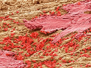
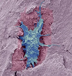
"Tissue: A Microscopic World Unveiled" Uterus lining during menstruation, SEM: Witness the intricate dance of life as the uterus sheds its lining during menstruation


All Professionally Made to Order for Quick Shipping
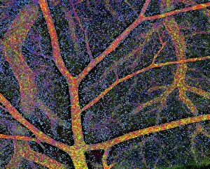
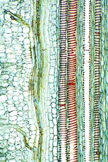
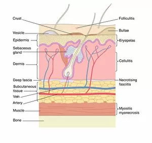
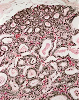
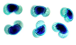
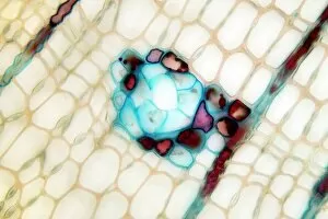
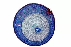
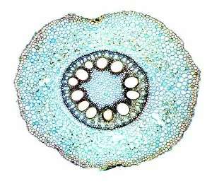
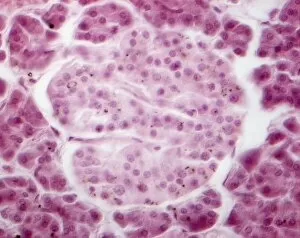
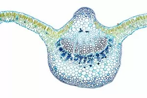
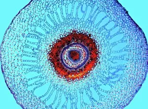
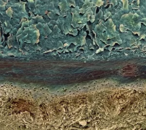
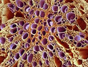
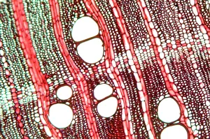
"Tissue: A Microscopic World Unveiled" Uterus lining during menstruation, SEM: Witness the intricate dance of life as the uterus sheds its lining during menstruation, revealing the resilience and beauty within. Dicotyledon plant stem, light micrograph: Delve into the mesmerizing patterns etched on a dicotyledon plant stem under a microscope's gaze, showcasing nature's artistry in every fiber. Skin disorders, artwork: Explore an artistic interpretation of skin disorders that unveils the hidden struggles faced by individuals while highlighting their strength and resilience. Brain tissue blood supply: Uncover the lifeline of our thoughts and emotions as we delve into the intricate network of blood vessels supplying oxygen to brain tissues - a testament to our cognitive prowess. Botanik Digitalis purpurea L. Fingerhut 160: 1: Immerse yourself in the captivating world of Botanik Digitalis purpurea L. , where delicate petals intertwine with vibrant hues, offering solace amidst nature's grandeur. Lactating breast tissue, light micrograph: Marvel at motherhood's miracle through a gentle lens capturing lactating breast tissue – an exquisite portrayal of nourishment and love personified. Blood cells: Embark on a journey within our veins as we explore blood cells bustling with life; each cell holds secrets that unlock vital clues about our health and well-being. Pine pollen grains, light micrograph: Peer into nature's microscopic marvels as pine pollen grains come alive under magnification – tiny particles carrying life across vast distances in pursuit of pollination perfection. Pine stem, light micrograph: Discover the sturdy elegance concealed beneath rough bark as you witness a pine stem reveal its inner intricacies through delicate beams of light – strength rooted deep within nature itself. Lime tree stem, light micrograph.