Fraud Blocker Skip to main content

False Colour Collection

"Unveiling the Hidden Beauty: Exploring False Colour in Microscopy and Imaging" Step into a world where reality meets artistry

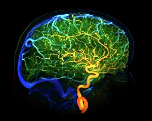
Background imageFalse Colour Collection: Brain blood vessels, 3D angiogram C007 / 1981

Brain blood vessels, 3D angiogram C007 / 1981
Brain blood vessels. Coloured 3D lateral angiogram (blood vessel X-ray) of the blood vessels in the brain of a healthy 32-year-old

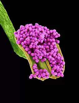
Background imageFalse Colour Collection: Geranium anther, SEM

Geranium anther, SEM
Geranium anther. Coloured scanning electron micrograph (SEM) of pollen (pink) on the anther of a geranium flower (Geranium sp.)

Background imageFalse Colour Collection: Dahlia flower pollen, SEM

Dahlia flower pollen, SEM
Dahlia flower pollen. Coloured scanning electron micrograph (SEM) of a pollen grain (orange) from a Dahlia cultorum flower. A pollen grain contains a male sex cell from a flowering plant

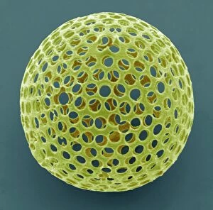
Background imageFalse Colour Collection: Acrosphaera radiolarian, SEM

Acrosphaera radiolarian, SEM
Acrosphaera radiolarian. Coloured scanning electron micrograph (SEM) of the shell of a Acrosphaera sp. radiolarian. Radiolaria are single-celled protozoans that are found in marine plankton

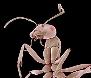
Background imageFalse Colour Collection: Ant, SEM

Ant, SEM
Ant. Coloured scanning electron micrograph (SEM) of an ant (family Formicidae). One of its compound eyes (red) is seen, as well as two long antennae that are mounted on the head between the eyes

Background imageFalse Colour Collection: Rough endoplasmic reticulum, TEM

Rough endoplasmic reticulum, TEM
Rough endoplasmic reticulum, coloured transmission electron micrograph (TEM). This section shows the rough endoplasmic reticulum (ER, folds, centre), a membranous structure that occurs in cells

Background imageFalse Colour Collection: Total hip replacement, X-ray

Total hip replacement, X-ray
Total hip replacement. Coloured frontal X-ray of a female pelvis with a total hip replacement (white, lower right). The replacement hip consists of a ball and shaft

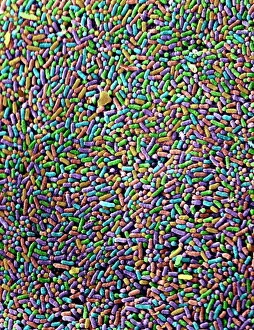
Background imageFalse Colour Collection: Salmonella bacteria, SEM

Salmonella bacteria, SEM
Salmonella bacteria, coloured scanning electron micrograph (SEM). Each rod-shaped structure is an individual bacterium. These bacteria occur mainly in human and animal intestines

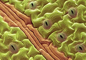
Background imageFalse Colour Collection: Leaf pores, SEM

Leaf pores, SEM
Leaf pores. Coloured scanning electron micrograph (SEM) of stomata (holes) on the surface of a leaf. These structures perform a similar function to the pores in human skin

Background imageFalse Colour Collection: Diatom, SEM

Diatom, SEM
Diatom. Coloured scanning electron micrograph (SEM) of a Stephanopyxis sp. diatom. Diatoms are single-celled photosynthetic algae, of which there are about 100, 000 species

Background imageFalse Colour Collection: Diatom, SEM

Diatom, SEM
Diatom. Coloured scanning electron micrograph (SEM) of a Triceratium morlandii diatom. Diatoms are single-celled photosynthetic algae, of which there are about 100, 000 species

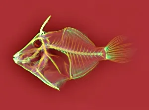
Background imageFalse Colour Collection: Triggerfish skeleton, X-ray

Triggerfish skeleton, X-ray
Triggerfish skeleton. Coloured X-ray of a triggerfish (family Balistidae). Its name originates from its ability to erect the first two dorsal spines (upper left), which can then lock or unlock

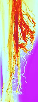
Background imageFalse Colour Collection: Varicose veins, X-ray

Varicose veins, X-ray
Varicose veins. Coloured X-ray of varicose veins in the calf of a fifty-five-year-old male. The veins (red), which carry blood back to the heart, are swollen, irregular and distorted

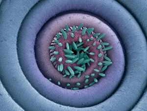
Background imageFalse Colour Collection: Moth proboscis, SEM

Moth proboscis, SEM
Moth proboscis. Coloured scanning electron micrograph (SEM) of the coiled proboscis of a moth (order Lepidoptera). The proboscis is an elongated part of the mouth

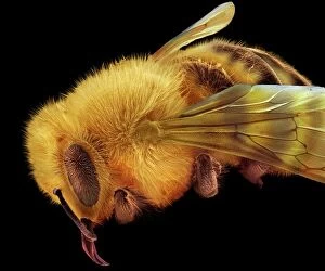
Background imageFalse Colour Collection: Honey bee, SEM

Honey bee, SEM
Honey bee (Apis mellifera), coloured scanning electron micrograph (SEM). The head and one of the two compound eyes is at lower left

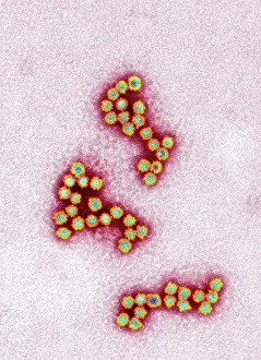
Background imageFalse Colour Collection: Norovirus particles, TEM

Norovirus particles, TEM
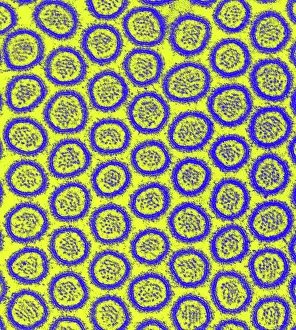
Norovirus particles. Coloured transmission electron micrograph (TEM) of norovirus particles. Norovirus is a genus of RNA (ribonucleic acid)

Background imageFalse Colour Collection: Bacteria infecting a macrophage, SEM

Bacteria infecting a macrophage, SEM
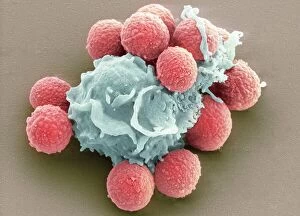
Bacteria infecting a macrophage. Coloured scanning electron micrograph (SEM) of Mycobacterium tuberculosis bacteria (purple) infecting a macrophage white blood cell

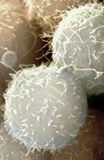
Background imageFalse Colour Collection: Stem cells, SEM

Stem cells, SEM
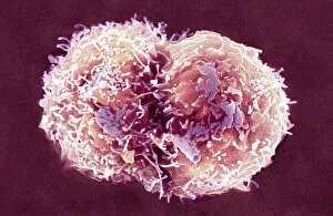
Stem cells, coloured scanning electron micrograph (SEM). Stem cells can differentiate into any other cell type. There are three main types of mammalian stem cell: embryonic stem cells

Background imageFalse Colour Collection: Geranium pollen, SEM

Geranium pollen, SEM
Geranium pollen. Coloured scanning electron micrograph (SEM) of pollen (pink) on the stamen of a geranium flower (Geranium sp.)

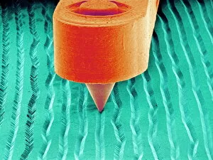
Background imageFalse Colour Collection: Needle playing a record, SEM

Needle playing a record, SEM
Needle playing a record. Coloured scanning electron micrograph (SEM) of the needle (stylus) of a record player in a groove on a record. A record is used to store sound

Background imageFalse Colour Collection: Medical zodiac, 15th century diagram

Medical zodiac, 15th century diagram
Medical zodiac. 15th century diagram with Latin text illustrating how the human body relates to the zodiac signs. Such information was used in medical practices, such as bloodletting

Background imageFalse Colour Collection: Macrophage engulfing TB bacteria, SEM

Macrophage engulfing TB bacteria, SEM
Macrophage engulfing TB bacteria. Coloured scanning electron micrograph (SEM) of a macrophage white blood cell (purple) engulfing a tuberculosis (Mycobacterium tuberculosis) bacterium (pink)

Background imageFalse Colour Collection: Diatom frustule, SEM

Diatom frustule, SEM
Diatom frustule. Coloured scanning electron micrograph (SEM) of the frustule of a Pinnularia sp. diatom. The diatoms are a group of photosynthetic, single-celled algae containing about 10

Background imageFalse Colour Collection: Head of a young newt, SEM

Head of a young newt, SEM
Head of a young newt. Scanning electron micrograph (SEM) of a juvenile (immature) newt. Although this newt is in the last stages of development

Background imageFalse Colour Collection: Phagocytosis of fungal spores, SEM

Phagocytosis of fungal spores, SEM
Phagocytosis of a fungus spore. Coloured scanning electron micrograph (SEM) of a neutrophil (blue) phagocytosing (engulfing and destroying) spores from the fungus Aspergillus fumigatus (red)

Background imageFalse Colour Collection: Neutrophil cell trapping bacteria, SEM

Neutrophil cell trapping bacteria, SEM
Neutrophil cell trapping bacteria. Coloured scanning electron micrograph (SEM) of bacteria (rod-shaped) being trapped by a neutrophil cell

Background imageFalse Colour Collection: Cauliflower head, SEM

Cauliflower head, SEM
Cauliflower. Coloured scanning electron micrograph (SEM) of the head of a cauliflower (Brassica oleracea botrytis). The arrangement of florets around the head is according to a Fibonacci series

Background imageFalse Colour Collection: Blood clot, SEM

Blood clot, SEM
Blood clot, coloured scanning electron micrograph (SEM). Red blood cells are red and fibrin protein strands are green. Platelets are at bottom right

Background imageFalse Colour Collection: Intestinal microvilli, TEM

Intestinal microvilli, TEM
Intestinal microvilli, coloured transmission electron micrograph (TEM). The microvilli are finger-like projections (seen here in circular cross-section)

Background imageFalse Colour Collection: Dinoflagellate plankton, SEM

Dinoflagellate plankton, SEM
Dinoflagellate plankton. Coloured scanning electron micrograph (SEM) of the calcareous (calcium-based) test (shell) of a dinoflagellate Calciodinellum sp

Background imageFalse Colour Collection: Spiny spider, SEM

Spiny spider, SEM
Spiny spider. Coloured scanning electron micrograph (SEM) of a spiny spider (Gasteracantha sp.), showing its characteristic spines (red) and large mouthparts (grey, centre left)

Background imageFalse Colour Collection: Calcium phosphate crystal, SEM

Calcium phosphate crystal, SEM
Calcium phosphate crystal, coloured scanning electron micrograph (SEM). Crystalline materials have their atoms placed in regular lattices that can form geometric shapes like this

Background imageFalse Colour Collection: Bacteria infecting a macrophage, SEM

Bacteria infecting a macrophage, SEM
Bacteria infecting a macrophage. Coloured scanning electron micrograph (SEM) of Mycobacterium tuberculosis bacteria (purple) infecting a macrophage white blood cell

Background imageFalse Colour Collection: Leaf oil glands, SEM

Leaf oil glands, SEM
Leaf oil glands. Coloured scanning electron micrograph (SEM) of a thyme plants leaf surface (Thymus vulgaris). Two types of leaf trichomes (hairs) are seen

Background imageFalse Colour Collection: Pollen on a bee leg, SEM

Pollen on a bee leg, SEM
Pollen on a bee leg. Coloured scanning electron micrograph (SEM) of pollen grains (pale purple) on the leg of a bee (superfamily Apoidea). Pollen grains are the male sex cells of a flowering plant

Background imageFalse Colour Collection: Dinoflagellates, SEM

Dinoflagellates, SEM
Dinoflagellates. Coloured scanning electron micrograph (SEM) of Prorocentrum dinoflagellates (round) and Ceratium dinoflagellates (branched). Dinoflagellates are unicellular protozoans

Background imageFalse Colour Collection: Red blood cells, SEM

Red blood cells, SEM
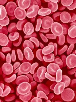
Red blood cells. Coloured scanning electron micrograph (SEM) of red blood cells (erythrocytes). Red blood cells are biconcave, disc-shaped cells that transport oxygen from the lungs to body cells

Background imageFalse Colour Collection: Pine tree stem, light micrograph

Pine tree stem, light micrograph
Pine tree stem. Coloured light micrograph of a cross-section through the stem of a pine tree. (Pinus sp.). The red wood (xylem, lower left) has resin canals in it, which are outlined in green

Background imageFalse Colour Collection: Diatom cell wall, SEM

Diatom cell wall, SEM
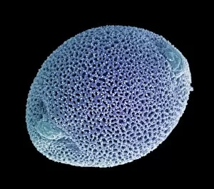
Diatom cell wall. Coloured scanning electron micrograph (SEM) of the pattern of small holes, called striae, in the cell wall of a diatom alga. This is a Coscinodiscus sp. diatom

Background imageFalse Colour Collection: Nerve synapse, TEM

Nerve synapse, TEM
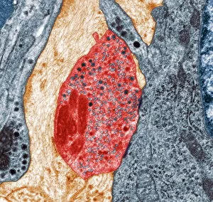
Nerve synapse. Coloured transmission electron micrograph (TEM) of the neuron (nerve) terminal at a synapse in the diaphragm

Background imageFalse Colour Collection: Stem cell, SEM

Stem cell, SEM
Stem cell, coloured scanning electron micrograph (SEM). Stem cells can differentiate into any other cell type. There are three main types of mammalian stem cell: embryonic stem cells

Background imageFalse Colour Collection: Stem cells, SEM

Stem cells, SEM
Stem cells, coloured scanning electron micrograph (SEM). Stem cells can differentiate into any other cell type. There are three main types of mammalian stem cell: embryonic stem cells

Background imageFalse Colour Collection: Normal torso, MRI scan

Normal torso, MRI scan
Normal torso, coloured profile (sagittal) magnetic resonance imaging (MRI) scan. The front of the body is at left. The spinal bones (vertebrae)

Background imageFalse Colour Collection: Chickweed pollen grains, SEM

Chickweed pollen grains, SEM
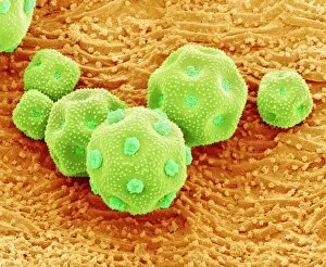
Chickweed pollen grains. Coloured scanning electron micrograph (SEM) of pollen grains (green) from a chickweed flower (Stellaria media)

Background imageFalse Colour Collection: Flame lily pollen, SEM

Flame lily pollen, SEM
Flame lily (Gloriosa sp.) pollen grains, coloured scanning electron micrograph (SEM). The grains have a highly sculptured outer wall (exine)

Background imageFalse Colour Collection: Xylem tissue, SEM

Xylem tissue, SEM
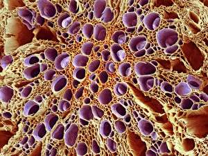
Xylem tissue. Coloured scanning electron micrograph (SEM) of a section through xylem tissue from a dicotyledon rootlet. Xylem vessels (purple)

Background imageFalse Colour Collection: Flower, SEM

Flower, SEM
Flower. Coloured scanning electron micrograph (SEM) of a flower

Background imageFalse Colour Collection: Spider lily flower stamen, SEM

Spider lily flower stamen, SEM
Spider lily flower stamen. Coloured scanning electron micrograph (SEM) of a cilia (hair-like structures) from a spider lily flower (Hymenocallis sp.) stamen

Background imageFalse Colour Collection: Daisy bud, SEM

Daisy bud, SEM
Daisy. Coloured scanning electron micrograph of a daisy bud (family Asteraceae, formerly Compositae). The protective bracts (green) cover clusters of tiny flowers, or florets

Background imageFalse Colour Collection: Zinnia flower petal, SEM

Zinnia flower petal, SEM
Zinnia flower petal. Coloured scanning electron micrograph (SEM) of a Zinnia sp. petal. The projections on the petal help to prevent water loss

Background imageFalse Colour Collection: Pollination, SEM

Pollination, SEM
Pollination. Coloured scanning electron micrograph (SEM) of part of a flowers pistil with pollen grains attached. The pistil is the female reproductive part of a flower

Background imageFalse Colour Collection: Haematopoietic stem cells, SEM C013 / 5009

Haematopoietic stem cells, SEM C013 / 5009
Haematopoietic stem cells, coloured scanning electron micrograph (SEM). Stem cells can differentiate into any other cell type

Background imageFalse Colour Collection: Human chromosomes, SEM C013 / 5002

Human chromosomes, SEM C013 / 5002
Human chromosomes. Coloured scanning electron micrograph (SEM) of human chromosomes prepared with the harlequin staining technique

Background imageFalse Colour Collection: Osteocyte, SEM

Osteocyte, SEM
Osteoblast bone cell. Coloured scanning electron micrograph (SEM) of an osteoblast bone cell. Osteocytes are osteoblasts (bone-producing cells) that have become trapped within bone cavities (lacunae)

Background imageFalse Colour Collection: Phagocytosis of fungus spores, SEM

Phagocytosis of fungus spores, SEM
Phagocytosis of fungus spores. Coloured scanning electron micrograph (SEM) of spores from the fungus Aspergillus fumigatus (red) being phagocytosed (engulfed and destroyed) by a dendritic cell (brown)

Background imageFalse Colour Collection: Plant cell mitosis, light micrograph

Plant cell mitosis, light micrograph
Plant cell mitosis. Light micrograph of root tip cells from an onion (Allium sp.) undergoing cell division (mitosis). From top left to bottom right

Background imageFalse Colour Collection: Monkey skeleton

Monkey skeleton
Marmoset skeleton. Coloured X-ray of a marmoset monkey (genus Callithrix) specimen. The monkey is held in place by wire supports (white vertical lines) which are attached to a base (bottom)

Background imageFalse Colour Collection: Sheep skull

Sheep skull. Coloured X-ray of a sagittal section through a sheeps (Ovis aries) skull. Sheep have 32 permanent teeth; 12 molars and eight incisors on the bottom jaw and 12 molars on the top jaw

Background imageFalse Colour Collection: Turtle ants head, SEM

Turtle ants head, SEM
Turtle ants head. Coloured scanning electron micrograph (SEM) of the head of a soldier turtle ant (Cephalotes sp.) from the Amazonian rainforest. One of its compound eyes (green) is at left

Background imageFalse Colour Collection: Nose mucosa, light micrograph

Nose mucosa, light micrograph
Nose mucosa. Light micrograph of a section through the nasal mucosa (the tissue that lines the airways of the nose), in the region lying over the nasal concha

Background imageFalse Colour Collection: Bone tissue

Bone tissue. Coloured scanning electron micrograph (SEM) of cancellous (spongy) bone. Bone tissue can be either cortical (compact) or cancellous

Background imageFalse Colour Collection: Metal shavings

Metal shavings. Coloured scanning electron micrograph (SEM) of metal shavings taken from a key cutter s

Background imageFalse Colour Collection: Medical zodiac, 15th century diagram

Medical zodiac, 15th century diagram
Medical zodiac. 15th century diagram with Greek text illustrating how the human body relates to the zodiac signs. Such information was used in medical practices, such as bloodletting

Background imageFalse Colour Collection: Aortic dissection, 3D CT scan

Aortic dissection, 3D CT scan
Aortic dissection. 3D computed tomography (CT) scan of a 67-year-old man with an aortic dissection. The aorta, the bodys main artery, is running vertically down centre (red)

Background imageFalse Colour Collection: Normal head and brain, MRI scan

Normal head and brain, MRI scan
Normal head and brain. Coloured profile (sagittal) magnetic resonance imaging (MRI) scan through the centre of the head and brain

Background imageFalse Colour Collection: Osteoblast bone cell, SEM

Osteoblast bone cell, SEM
Osteoblast bone cell. Coloured scanning electron micrograph (SEM) of an osteoblast (centre) in bone tissue. Osteoblasts are primarily found in regions of new bone growth

Background imageFalse Colour Collection: Passion flower pollen, SEM

Passion flower pollen, SEM
Passion flower pollen. Coloured scanning electron micrograph (SEM) of pollen grains from a passion flower (Passiflora caerulea). Pollen grains are the male gametes (sex cells) of a plant

Background imageFalse Colour Collection: Smallpox virus particle, TEM

Smallpox virus particle, TEM
Smallpox virus particle. Coloured transmission electron micrograph (TEM) of a Variola major virus particle. Each particle consists of a DNA (deoxyribonucleic acid)

Background imageFalse Colour Collection: Infectious bronchitis virus (IBV), TEM

Infectious bronchitis virus (IBV), TEM
Infectious bronchitis virus (IBV) particles, coloured transmission electron micrograph (TEM). This virus is a member of the coronavirus family

Background imageFalse Colour Collection: Ant, SEM

Ant, SEM
Ant (family Formicidae), coloured scanning electron micrograph (SEM). One of its compound eyes (black) is seen, as well as two long antennae that are mounted on the head between the eyes

Background imageFalse Colour Collection: Rene Descartes and Isaac Beeckman

Rene Descartes and Isaac Beeckman. Historical engraving of the French mathematician & philosopher Rene Descartes (1596- 1650, centre)

Background imageFalse Colour Collection: Flakes of dead skin, SEM

Flakes of dead skin, SEM
Flakes of dead skin. Coloured scanning electron micrograph (SEM) of dead skin from the scalp (dandruff), a condition where there is an excessive shedding of dead skin cells

Background imageFalse Colour Collection: Chickweed pollen grain, SEM

Chickweed pollen grain, SEM
Chickweed pollen grain. Coloured scanning electron micrograph (SEM) of a pollen grain (green) from a chickweed flower (Stellaria media)

Background imageFalse Colour Collection: Root nodule

Root nodule. Coloured scanning electron micrograph (SEM) of a root nodule on a pea plant (Pisum sativum) caused by the nitrogen-fixing soil bacteria Rhizobium leguminosarum

Background imageFalse Colour Collection: Fungal spores, SEM

Fungal spores, SEM
Fungal spores. Coloured scanning electron micrograph (SEM) of fungal spores on a fruiting body of an Aspergillus sp. fungus. The name for such a fruiting body is a conidiophore

Background imageFalse Colour Collection: Bouncing ball

Bouncing ball, stroboscopic image using coloured lights. The strobe light was set to flash 50 times every second, and the exposure time was about 1.5 seconds

Background imageFalse Colour Collection: Credit Image courtesy: MODIS Ocean Group, NASA GSFC, and the University of Miami

Credit Image courtesy: MODIS Ocean Group, NASA GSFC, and the University of Miami
Credit Image courtesy:MODIS Ocean Group, NASA GSFC, and the University of Miami: sea surface temperature shown in false-colour image. a one-month composite for May 2001

Background imageFalse Colour Collection: false-colour images feature the Indus River Valley, a lush oasis of vegetation made

false-colour images feature the Indus River Valley, a lush oasis of vegetation made possible by the Indus River, which is visible as a thin black thread heading toward the Arabian Sea in

Background imageFalse Colour Collection: Spitzer Space Telescope false-colour image of South Pillar region of tsar-forming

Spitzer Space Telescope false-colour image of South Pillar region of tsar-forming region called the Carina Nebula. Star embryos (yellow or white) inside finger-like pillars of thick dust (pink)

Background imageFalse Colour Collection: False-colour image of supernova remnant Cassiopeia A in constellation Cassiopeia

False-colour image of supernova remnant Cassiopeia A in constellation Cassiopeia, 10, 000 light-years away. It is the remnant of a once massive star that died in a supernova explosion. Credit NASA

Background imageFalse Colour Collection: Spitzer Space Telescope false-colour image of mountains where stars are born

Spitzer Space Telescope false-colour image of mountains where stars are born. Dubbed Mountains of Creation the towering pillars of cool gas and dust are lit at the tips with light from embryo stars

Background imageFalse Colour Collection: Optical light microscope with camera

Optical light microscope with camera
Microscope. Computer artwork of a typical optical microscope with a digital camera attached for photography. Below the camera these are the two eyepieces (binocular) for viewing the specimen

Background imageFalse Colour Collection: Optical light microscope, artwork

Optical light microscope, artwork
Microscope. Computer artwork of a typical optical microscope. Below the camera these are the two eyepieces (binocular) for viewing the specimen

Background imageFalse Colour Collection: Saudi Arabia agriculture, 2000

Saudi Arabia agriculture, 2000
Saudi Arabia agriculture. False-colour satellite image showing the extent of agricultural operations in the Wadi As-Sirhan Basin of Saudi Arabia in 2000


All Professionally Made to Order for Quick Shipping

"Unveiling the Hidden Beauty: Exploring False Colour in Microscopy and Imaging" Step into a world where reality meets artistry, as we delve into the captivating realm of "false colour. " In this mesmerizing journey, we encounter an array of intricate subjects that have been transformed through advanced imaging techniques. Brace yourself for a visual feast that transcends our conventional perception. First up, behold the brain's intricate network of blood vessels captured in a 3D angiogram from 1981. Through false colour enhancement, these delicate pathways come alive with vibrant hues, revealing their hidden complexity. Moving on to nature's wonders, prepare to be enchanted by the Geranium anther under scanning electron microscopy (SEM). Its microscopic details are unveiled in vivid shades, showcasing its remarkable structure and beauty. Next on our exploration is Dahlia flower pollen also observed under SEM. The once minuscule grains now emerge as magnificent orbs adorned with striking colours—a testament to nature's exquisite design. Venturing further into the microscopic realm, we encounter diatoms—tiny algae—with their intricately patterned shells. Under SEM and enhanced with false colouring techniques, these diatoms transform into kaleidoscopic masterpieces that blur the line between science and art. But it doesn't stop there. Witness an ant like never before through SEM; its exoskeleton becomes a work of art when illuminated by vibrant tones. Meanwhile, X-ray imaging reveals the intricate skeleton of a triggerfish—an ethereal display reminiscent of otherworldly creatures lurking beneath ocean depths. Delving deeper within cells brings us face-to-face with rough endoplasmic reticulum—the cellular factory responsible for protein synthesis. Enhanced by false colouring using transmission electron microscopy (TEM), this complex network takes on an otherworldly appearance akin to futuristic cityscapes bathed in neon lights. Shifting gears towards medical marvels captured via X-ray technology unveils both the functional and aesthetic aspects of our bodies.

© Copyright Media Storehouse, All Rights Reserved 2003 - 2024
Facebook     Pinterest     Card Payments     Reviews IO