Fraud Blocker Skip to main content

False Colour Collection (page 5)

"Unveiling the Hidden Beauty: Exploring False Colour in Microscopy and Imaging" Step into a world where reality meets artistry

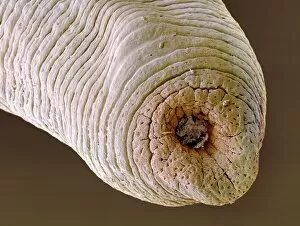
Background imageFalse Colour Collection: Freshwater leechs head sucker, SEM

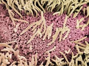
Freshwater leechs head sucker, SEM
Freshwater leechs head sucker. Coloured scanning electron micrograph (SEM) of the head sucker of a freshwater leech (Hirudo sp.). This parasite has two suckers

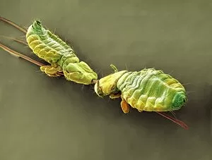
Background imageFalse Colour Collection: Badger body lice, SEM

Badger body lice, SEM
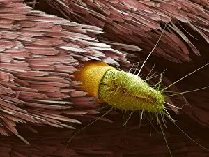
Badger body lice. Coloured scanning electron micrograph (SEM) of two badger body lice (Trichodectes melis) on badger hairs

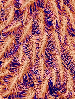
Background imageFalse Colour Collection: Crab feeding structures, SEM

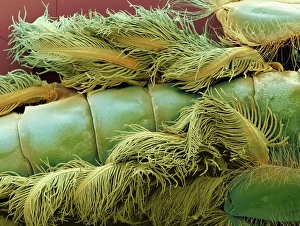
Crab feeding structures, SEM
Crab filter-feeding structures. Coloured scanning electron micrograph (SEM) of filtering structures (setae) that are part of the feeding structures of a crab (order Decapoda)

Background imageFalse Colour Collection: Replica machine-gun

Replica machine-gun
Replica machine gun

Background imageFalse Colour Collection: Anglers swivel, SEM

Anglers swivel, SEM
Swivel, coloured scanning electron micrograph (SEM). This is used by anglers to prevent tangles in their lines while fishing

Background imageFalse Colour Collection: Lead-free angling shot, SEM

Lead-free angling shot, SEM
Lead-free angling Shot, coloured scanning electron micrograph (SEM). This metal ball is used by anglers to weight fishing line. Unlike older weights, this shot contains no lead

Background imageFalse Colour Collection: Moths interior, SEM

Moths interior, SEM
Moths interior. Coloured scanning electron micrograph (SEM) of the trachea and Malpighi tubules of a moth (order Lepidoptera)

Background imageFalse Colour Collection: Squirrel flea antenna, SEM

Squirrel flea antenna, SEM
Squirrel flea antenna. Coloured scanning electron micrograph (SEM) of the antenna of a Squirrel flea (Orchopeas howardi howardi)

Background imageFalse Colour Collection: Hedgehog tick, SEM

Hedgehog tick, SEM
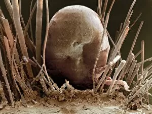
Hedgehog tick. Coloured scanning electron micrograph (SEM) of a hedgehog tick (Ixodes hexagonus) feeding. Its body is swollen with blood. Ticks are parasitic arachnids

Background imageFalse Colour Collection: Scorpion fly head, SEM

Scorpion fly head, SEM
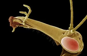
Scorpion fly head. Coloured scanning electron micrograph (SEM) of the head of a scorpion fly (Panorpa sp.). It has a long beak-like extension on its head that has the jaws at the end (left)

Background imageFalse Colour Collection: Lacewing larva, SEM

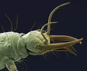
Lacewing larva, SEM
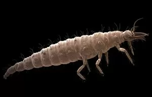
Lacewing larva (Chrysoperla carnea), coloured scanning electron micrograph (SEM). This insect preys on aphids, whitefly and other insects

Background imageFalse Colour Collection: Geometer moth, SEM

Geometer moth, SEM
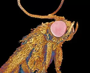
Geometer moth. Coloured scanning electron micrograph (SEM) of a moth belonging to the family Geometridae. The head is dominated by two large compound eyes (red)

Background imageFalse Colour Collection: Bee mites, SEM

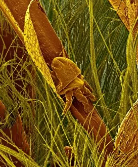
Bee mites, SEM
Bee mites. Coloured scanning electron micrograph (SEM) of bee mites on the body of a bee. Some bee mite species feed by cutting into the surface membranes of the bee

Background imageFalse Colour Collection: Lacewing larva head, SEM

Lacewing larva head, SEM
Lacewing larva head (Chrysoperla carnea), coloured scanning electron micrograph (SEM). This insect preys on aphids, whitefly and other insects

Background imageFalse Colour Collection: Crown-of-thorns starfish skeleton, X-ray

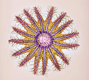
Crown-of-thorns starfish skeleton, X-ray
Crown-of-thorns starfish skeleton. Coloured X-ray of the crown-of-thorns starfish (Acanthaster planci). This starfish lives on coral reefs in the Indo-Pacific region

Background imageFalse Colour Collection: Tarantula foot, SEM

Tarantula foot, SEM
Tarantula foot. Coloured scanning electron micrograph (SEM) of a part of a tarantulas foot. The two claws are used to grip rough surfaces

Background imageFalse Colour Collection: Centipede underside, SEM

Centipede underside, SEM
Centipede. Coloured scanning electron micrograph (SEM) of the underside of a centipede. Centipedes (class Chilopoda) are arthropods with elongated bodies and one pair of legs per segment

Background imageFalse Colour Collection: Gills of an asian conch

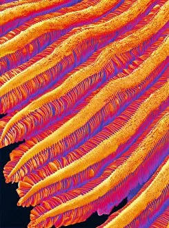
Gills of an asian conch
Gills. Coloured scanning electron micrograph (SEM) of the gills of an asian conch (family Viviparidae), also known as a mystery snail

Background imageFalse Colour Collection: Bee mite, SEM

Bee mite, SEM
Bee mite. Coloured scanning electron micrograph (SEM) of a bee mite on a bee. Some bee mite species feed by cutting into the surface membranes of the bee

Background imageFalse Colour Collection: Underside of a slug, SEM

Underside of a slug, SEM
Slug. Coloured scanning electron micrograph (SEM) of the underside of a slug (order Gastropoda). The underside of the slug is covered in microscopic hair-like projections known as cilia (yellow)

Background imageFalse Colour Collection: Moth antennae, SEM

Moth antennae, SEM
Moth antennae. Coloured scanning electron micrograph (SEM) of the antennae of a moth (order Lepidoptera). The antennae are covered in sensory hairs that detect movement and smell

Background imageFalse Colour Collection: Jumping spiders leg, SEM

Jumping spiders leg, SEM
Jumping spiders leg (family Salticidae). Coloured scanning electron micrograph (SEM) of the tip of a jumping spiders leg. The hairs are used for gripping and also as a sensory tool

Background imageFalse Colour Collection: Squirrel flea, SEM

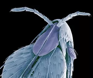
Squirrel flea, SEM
Squirrel flea (Orchopeas howardi howardi), coloured scanning electron micrograph (SEM). The two structures protruding from the centre of the fleas head are sensory palps

Background imageFalse Colour Collection: Pond snails, SEM

Pond snails, SEM
Pond snails. Coloured scanning electron micrograph (SEM) of the shells of snails. These snails live in ponds. Snails are molluscs that protect themselves with a mineralised spiralling shell

Background imageFalse Colour Collection: Honey bee mite, SEM

Honey bee mite, SEM
Honey bee mite. Coloured scanning electron micrograph (SEM) of a honey bee mite (Varroa sp.). Parasitic mites have decimated hives of wild and domesticated honey bees (Apis mellifera)

Background imageFalse Colour Collection: Chrysalis, SEM

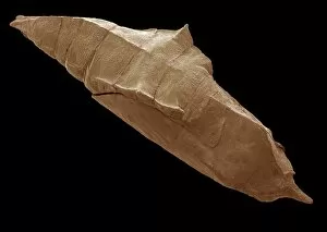
Chrysalis, SEM
Chrysalis, coloured scanning electron micrograph (SEM). A chrysalis is the name for the pupa of butterflies and moths (order Lepidoptera)

Background imageFalse Colour Collection: Gills of an Alderfly larva, SEM

Gills of an Alderfly larva, SEM
Gills of an alderfly larva. Coloured scanning electron micrograph (SEM) of the gills of an alderfly (Sialis lutaria) larva. The gills are the feather-like structures seen at top and bottom

Background imageFalse Colour Collection: Tamarins skin

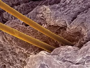
Tamarins skin. Scanning electron micrograph of a tamarins (Saguinus sp.) skin, with two hairs (yellow) emerging from it. Tamarins are a small type of New World monkey

Background imageFalse Colour Collection: Hedgehog skin and spines, SEM

Hedgehog skin and spines, SEM
Hedgehog skin and spines. Coloured scanning electron micrograph (SEM) of the skin of a hedgehog, with spines (blue) protruding from it. A spine is a modified hair made from the protein keratin

Background imageFalse Colour Collection: Tamarins skin and hairs

Tamarins skin and hairs
Tamarins skin. Scanning electron micrograph of a tamarins (Saguinus sp.) skin, with hairs (brown) emerging from it. Tamarins are a small type of New World monkey

Background imageFalse Colour Collection: Striated finch skull, SEM

Striated finch skull, SEM
Striated finch skull. Coloured scanning electron micrograph (SEM) of the skull of the striated finch (Lonchura striata), seen from underneath

Background imageFalse Colour Collection: Young newt, SEM

Young newt, SEM
Young newt. Scanning electron micrograph (SEM) of a juvenile (immature) newt. Although this newt is in the last stages of development

Background imageFalse Colour Collection: Hairs on a geckos toe

Hairs on a geckos toe. Coloured scanning electron micrograph (SEM) showing the numerous hairs which cover the underside of a geckos feet

Background imageFalse Colour Collection: Lovebirds claw, SEM

Lovebirds claw, SEM
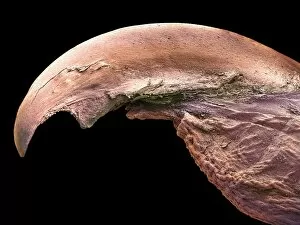
Lovebirds claw. Coloured scanning electron micrograph (SEM) of a claw (talon) on the toe of a Lovebird (Agapornis sp.). The claws and beaks of birds are made from keratin, a hard, fibrous protein

Background imageFalse Colour Collection: Mole nose, SEM

Mole nose, SEM
Mole nose. Coloured scanning electron micrograph (SEM) of the snout (pink) of a mole, showing specialised bulbous papillae (projections) known as Eimers organs

Background imageFalse Colour Collection: Underside of a geckos toe

Underside of a geckos toe. Coloured scanning electron micrograph (SEM) showing the numerous hairs which cover the underside of a geckos feet

Background imageFalse Colour Collection: Head of a young newt, SEM

Head of a young newt, SEM
Head of a young newt. Scanning electron micrograph (SEM) of a juvenile (immature) newt. Although this newt is in the last stages of development

Background imageFalse Colour Collection: Orangutan skull, X-ray

Orangutan skull, X-ray
Orangutan skull. Coloured X-ray of an orangutan (Pongo sp.) skull. The orangutan is native to Malaysia and Indonesia

Background imageFalse Colour Collection: Chimpanzee skull, X-ray

Chimpanzee skull, X-ray
Chimpanzee skull. Coloured X-ray of a side view of a chimpanzees (Pan troglodytes) skull. The cranium (upper right) is considerably smaller than in humans

Background imageFalse Colour Collection: Mole hair, SEM

Mole hair, SEM
Mole hair. Coloured scanning electron micrograph (SEM) of a skin of a mole (family Talpidae), showing fine hairs. Moles are small mammals that live in underground burrows

Background imageFalse Colour Collection: Orangutan skull

Orangutan skull. Coloured X-ray of an orangutan (Pongo sp.) skull. The orangutan is native to Malaysia and Indonesia

Background imageFalse Colour Collection: Rat skeleton

Rat skeleton. Coloured X-ray of a rat (Rattus sp.) specimen. The rat is held in place by wire supports (blue vertical lines) which are attached to a base (bottom)

Background imageFalse Colour Collection: Moth rear end, SEM

Moth rear end, SEM
Moth rear end. Coloured scanning electron micrograph (SEM) of the rear end of a moth (order Lepidoptera)


All Professionally Made to Order for Quick Shipping

"Unveiling the Hidden Beauty: Exploring False Colour in Microscopy and Imaging" Step into a world where reality meets artistry, as we delve into the captivating realm of "false colour. " In this mesmerizing journey, we encounter an array of intricate subjects that have been transformed through advanced imaging techniques. Brace yourself for a visual feast that transcends our conventional perception. First up, behold the brain's intricate network of blood vessels captured in a 3D angiogram from 1981. Through false colour enhancement, these delicate pathways come alive with vibrant hues, revealing their hidden complexity. Moving on to nature's wonders, prepare to be enchanted by the Geranium anther under scanning electron microscopy (SEM). Its microscopic details are unveiled in vivid shades, showcasing its remarkable structure and beauty. Next on our exploration is Dahlia flower pollen also observed under SEM. The once minuscule grains now emerge as magnificent orbs adorned with striking colours—a testament to nature's exquisite design. Venturing further into the microscopic realm, we encounter diatoms—tiny algae—with their intricately patterned shells. Under SEM and enhanced with false colouring techniques, these diatoms transform into kaleidoscopic masterpieces that blur the line between science and art. But it doesn't stop there. Witness an ant like never before through SEM; its exoskeleton becomes a work of art when illuminated by vibrant tones. Meanwhile, X-ray imaging reveals the intricate skeleton of a triggerfish—an ethereal display reminiscent of otherworldly creatures lurking beneath ocean depths. Delving deeper within cells brings us face-to-face with rough endoplasmic reticulum—the cellular factory responsible for protein synthesis. Enhanced by false colouring using transmission electron microscopy (TEM), this complex network takes on an otherworldly appearance akin to futuristic cityscapes bathed in neon lights. Shifting gears towards medical marvels captured via X-ray technology unveils both the functional and aesthetic aspects of our bodies.