Fraud Blocker Skip to main content

False Colour Collection (page 7)

"Unveiling the Hidden Beauty: Exploring False Colour in Microscopy and Imaging" Step into a world where reality meets artistry

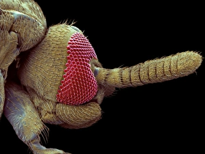
Background imageFalse Colour Collection: Yellow dung fly head, SEM

Yellow dung fly head, SEM
Yellow dung fly head. Coloured scanning electron micrograph (SEM) of the head of a yellow dung fly (Scatophaga stercoraria)

Background imageFalse Colour Collection: Gnat head, SEM

Gnat head, SEM
Gnat head. Coloured scanning electron micrograph (SEM) of the head of a gnat, a small type of fly (order Diptera), seen from the side

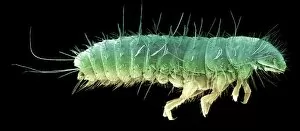
Background imageFalse Colour Collection: Bloodworm tail

Bloodworm tail
Bloodworms tail. Coloured scanning electron micrograph (SEM) of the tail of a bloodworm (Chironomus sp.). The bloodworm is the freshwater larva of a midge

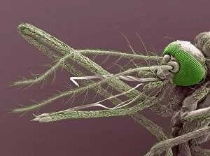
Background imageFalse Colour Collection: Mosquito mouthparts, SEM

Mosquito mouthparts, SEM
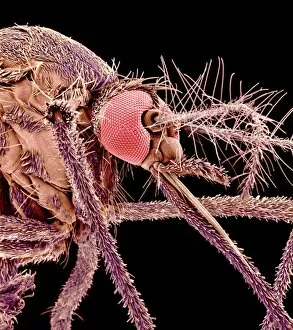
Mosquito mouthparts. Coloured scanning electron micrograph (SEM) of the mouthparts of a mosquito (family Culicidae). The head is at far right, with compound eyes (green)

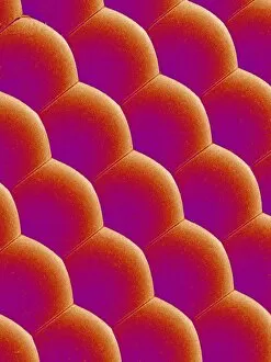
Background imageFalse Colour Collection: Compound eye of a mosquito, SEM

Compound eye of a mosquito, SEM
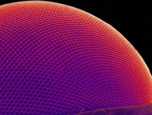
Compound eye of a mosquito. Coloured scanning electron micrograph (SEM) of numerous lenses making up the surface of a compound eye from an Asian tiger mosquito (Aedes albopictus)

Background imageFalse Colour Collection: Flys eye

Flys eye. Coloured scanning electron micrograph (SEM) of a flys (order Diptera) compound eye. The eye is made up of numerous visual units, known as ommatidia

Background imageFalse Colour Collection: Compound eye of a moth, SEM

Compound eye of a moth, SEM
Moths eye. Coloured scanning electron micrograph (SEM) of a section of a moths compound eye. The eye has been sliced to show its internal structure

Background imageFalse Colour Collection: Beetle larva, SEM

Beetle larva, SEM
Beetle larva. Coloured scanning electron micrograph (SEM) of a beetle larva (order Coleoptera)

Background imageFalse Colour Collection: Greenfly, SEM

Greenfly, SEM
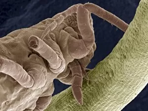
Greenfly. Coloured scanning electron micrograph (SEM) of an aphid (sub-order Homoptera), or greenfly, feeding on a plant stem (right, green)

Background imageFalse Colour Collection: Great diving beetles foreleg, SEM

Great diving beetles foreleg, SEM
Great diving beetles foreleg. Coloured scanning electron micrograph (SEM) of the foreleg of a male great diving beetle (Dytiscus marginalis). At the tip of the leg are two claws

Background imageFalse Colour Collection: Soldier ant jaws, SEM

Soldier ant jaws, SEM
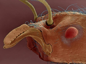
Soldier ant jaws. Coloured scanning electron micrograph (SEM) of the head of a leaf-cutter ant (Atta sexdens). This is a soldier ant

Background imageFalse Colour Collection: Breathing tube on a fruit flys pupa, SEM

Breathing tube on a fruit flys pupa, SEM
Breathing tube on a fruit flys pupa, coloured scanning electron micrograph (SEM). This structure is called a spiracle. This fruit fly is Drosophila melanogaster (wild type Oregon R)

Background imageFalse Colour Collection: Leaf cutter ant, SEM

Leaf cutter ant, SEM
Leaf cutter ants head. Coloured scanning electron micrograph (SEM) of the head of a leaf cutter ant. This insect lives in colonies and uses its powerful mandibles to remove pieces of leaves

Background imageFalse Colour Collection: Fly glandular organ, SEM

Fly glandular organ, SEM
Fly glandular organ. Coloured scanning electron micrograph (SEM) of a glandular organ on a March fly (family Bibionidae). This organ is found on the rear of the fly

Background imageFalse Colour Collection: Hercules beetle antenna, SEM

Hercules beetle antenna, SEM
Hercules beetle antenna. Coloured scanning electron micrograph (SEM) of the antenna of a hercules beetle (Dynastes hercules)

Background imageFalse Colour Collection: Moth antenna

Moth antenna. Coloured scanning electron micrograph (SEM) of the antenna of a moth (order Lepidoptera). The antenna is covered in sensory hairs that detect movement and smell

Background imageFalse Colour Collection: Mosquito antenna, SEM

Mosquito antenna, SEM
Mosquito antenna. Coloured scanning electron micrograph (SEM) of one of a mosquitos antennae. The antenna is covered with hairs that contain sensory cells

Background imageFalse Colour Collection: Insect foot, SEM

Insect foot, SEM
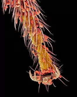
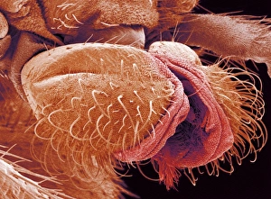
Insect foot. Coloured electron micrograph (SEM) of an unidentified insects foot. The foot consists of adhesive pads (red, upper left and right)

Background imageFalse Colour Collection: Fly compound eye, SEM

Fly compound eye, SEM
Fly compound eye. Coloured scanning electron micrograph (SEM) of numerous lenses making up the surface of a compound eye from a hover fly (family Syrphidae). These lenses are called ommatidia

Background imageFalse Colour Collection: Mosquito wing surface, SEM

Mosquito wing surface, SEM
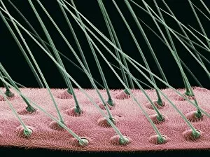
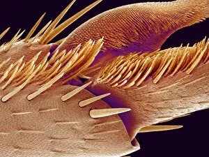
Mosquito wing surface. Coloured scanning electron micrograph (SEM) of the micro-structures on the surface of a mosquito (family Culicidae) wing

Background imageFalse Colour Collection: Tip of ant foot, SEM

Tip of ant foot, SEM
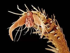
Tip of ant foot. Coloured scanning electron micrograph (SEM) of the tip of a leg from an ant (family Formicidae). The end of an insect leg consists of the final segment, which is called the tarsus

Background imageFalse Colour Collection: Ant foot, SEM

Ant foot, SEM
Ant foot. Coloured scanning electron micrograph (SEM) of the tip of a leg from an ant (family Formicidae). The end of an insect leg consists of the final segment, which is called the tarsus

Background imageFalse Colour Collection: Beetle antenna, SEM

Beetle antenna, SEM
Beetle antenna. Coloured scanning electron micrograph (SEM) of the antenna of a Fregate beetle (Polposipus herculeanus). The antenna is covered in sensory hairs and cells (yellow)

Background imageFalse Colour Collection: Moth proboscis

Moth proboscis. Coloured scanning electron micrograph (SEM) of the labial palps, which are found on either side of the proboscis (tubular feeding organ)

Background imageFalse Colour Collection: Carpet beetle larva hairs, SEM

Carpet beetle larva hairs, SEM
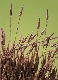
Beetle larva hairs. Coloured scanning electron micrograph (SEM) of hairs from a carpet beetle larva (family Dermestidae). These spear-headed hairs can often cause irritation when they come into

Background imageFalse Colour Collection: Carpet beetle larva hair, SEM

Carpet beetle larva hair, SEM
Beetle larva hair. Coloured scanning electron micrograph (SEM) of a hair from a carpet beetle larva (family Dermestidae). Large numbers of these spear-headed hairs can cause irritation when they come

Background imageFalse Colour Collection: Fly balance organ, SEM

Fly balance organ, SEM
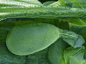
Fly balance organ. Coloured scanning electron micrograph (SEM) of the balance organ (haltere) of a fly (order Diptera). The haltere is the oval structure at centre and lower left

Background imageFalse Colour Collection: Long-horned beetle, SEM

Long-horned beetle, SEM
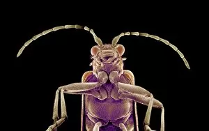
Long-horned beetle (family Cerambycidae), coloured scanning electron micrograph (SEM). Beetles in this family are characterized by very long antennae

Background imageFalse Colour Collection: Moth fly, SEM

Moth fly, SEM
Moth fly. Coloured scanning electron micrograph (SEM) of a moth fly (Psychoda sp.). The two compound eyes (pink) dominate the head

Background imageFalse Colour Collection: Mosquito heads, light micrograph

Mosquito heads, light micrograph
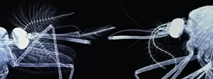
Mosquito heads. Light micrograph of the heads of a male (left) and female (right) mosquito (family Culicidae). The mosquitos head is dominated by its large compound eyes (solid white)

Background imageFalse Colour Collection: Thrips, SEM

Thrips, SEM
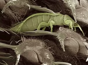
Thrips. Coloured scanning electron micrograph (SEM) of a thrips, or thunder fly (order Thysanoptera), feeding inside a grass seed head

Background imageFalse Colour Collection: Mosquito larva mouth hairs

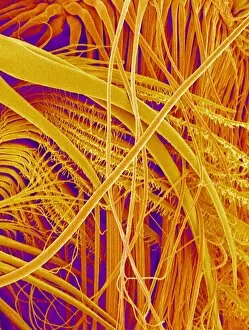
Mosquito larva mouth hairs. Coloured scanning electron micrograph (SEM) of the mouth hairs of a mosquito larva (Aedes albopictus). These hairs are feeding structures used to filter water

Background imageFalse Colour Collection: Devils coach horse beetle, SEM

Devils coach horse beetle, SEM
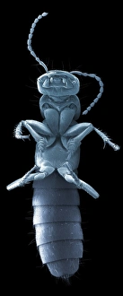
Devils coach horse. Coloured scanning electron micrograph (SEM) of the underside of a devils coach horse beetle (Staphylinus olens)

Background imageFalse Colour Collection: Fly foot, SEM

Fly foot, SEM
Fly foot. Coloured scanning electron micrograph (SEM) of the tip of a leg of a March fly (family Bibionidae). A fly uses claws (one seen here) to help it grip rough surfaces

Background imageFalse Colour Collection: Gnat foot, SEM

Gnat foot, SEM
Gnat foot. Coloured scanning electron micrograph (SEM) of the pads and claws on the tip of a leg of a gnat, a small fly (order Diptera)

Background imageFalse Colour Collection: Bee foot, SEM

Bee foot, SEM
Bee foot. Coloured scanning electron micrograph (SEM) of the tip of a leg from a bee (superfamily Apoidea). The end of an insect leg consists of the final segment, which is called the tarsus

Background imageFalse Colour Collection: Carpet beetle hair, SEM

Carpet beetle hair, SEM
Carpet beetle hair. Coloured scanning electron micrograph (SEM) of the spear-headed hair (hastisetae) of a carpet beetle larva (family Dermestidae)

Background imageFalse Colour Collection: Asian tiger mosquito, SEM

Asian tiger mosquito, SEM
Asian tiger mosquito (Aedes albopictus), coloured scanning electron micrograph (SEM). Structures on its head (upper centre) include its large compound eyes (red), two feather-like antennae (right)

Background imageFalse Colour Collection: Aphids, SEM

Aphids, SEM
Aphids. Coloured scanning electron micrograph (SEM) of aphids (family Aphididae), or greenflies, feeding on a plant stem. Only their abdomens can be seen here

Background imageFalse Colour Collection: Fly mouthparts, SEM

Fly mouthparts, SEM
Fly mouthparts. Coloured scanning electron micrograph (SEM) of the proboscis of a March fly (family Bibionidae). The proboscis is a feeding structure that is coiled up when not in use

Background imageFalse Colour Collection: Moth wing

Moth wing. Coloured scanning electron micrograph (SEM) of the wing of a moth (order Lepidoptera). A moth wing is covered in tiny scales that are modified hairs (setae)

Background imageFalse Colour Collection: Hercules beetle foot, SEM

Hercules beetle foot, SEM
Hercules beetle foot. Coloured scanning electron micrograph (SEM) of the foot of a hercules beetle (Dynastes hercules). The foot comprises a pair of hooked claws (centre)

Background imageFalse Colour Collection: Fungus weevil head, SEM

Fungus weevil head, SEM
Weevil head. Coloured scanning electron micrograph (SEM) of the head of an endangered fungus weevil (Notioxenus sp.). The mouthparts are at lower right

Background imageFalse Colour Collection: Moths interior

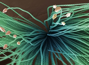
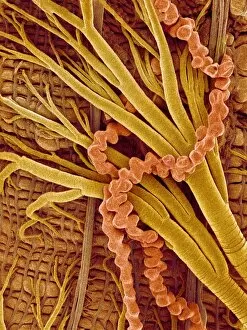
Moths interior. Scanning electron micrograph (SEM) of the trachea and Malpighi tubules of a moth (order Lepidoptera). Oxygen enters the tracheal tubes (green) from the spiracles (breathing pores)

Background imageFalse Colour Collection: Pupa, SEM

Pupa, SEM
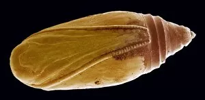
Pupa. Coloured scanning electron micrograph (SEM) of a Lepidopteran pupa

Background imageFalse Colour Collection: Ant with fungus, SEM

Ant with fungus, SEM
Ant with fungus. Coloured scanning electron micrograph (SEM) of a leaf-cutter ant (Atta sexdens) with a piece of fungus. This ant cuts leaves from foliage


All Professionally Made to Order for Quick Shipping

"Unveiling the Hidden Beauty: Exploring False Colour in Microscopy and Imaging" Step into a world where reality meets artistry, as we delve into the captivating realm of "false colour. " In this mesmerizing journey, we encounter an array of intricate subjects that have been transformed through advanced imaging techniques. Brace yourself for a visual feast that transcends our conventional perception. First up, behold the brain's intricate network of blood vessels captured in a 3D angiogram from 1981. Through false colour enhancement, these delicate pathways come alive with vibrant hues, revealing their hidden complexity. Moving on to nature's wonders, prepare to be enchanted by the Geranium anther under scanning electron microscopy (SEM). Its microscopic details are unveiled in vivid shades, showcasing its remarkable structure and beauty. Next on our exploration is Dahlia flower pollen also observed under SEM. The once minuscule grains now emerge as magnificent orbs adorned with striking colours—a testament to nature's exquisite design. Venturing further into the microscopic realm, we encounter diatoms—tiny algae—with their intricately patterned shells. Under SEM and enhanced with false colouring techniques, these diatoms transform into kaleidoscopic masterpieces that blur the line between science and art. But it doesn't stop there. Witness an ant like never before through SEM; its exoskeleton becomes a work of art when illuminated by vibrant tones. Meanwhile, X-ray imaging reveals the intricate skeleton of a triggerfish—an ethereal display reminiscent of otherworldly creatures lurking beneath ocean depths. Delving deeper within cells brings us face-to-face with rough endoplasmic reticulum—the cellular factory responsible for protein synthesis. Enhanced by false colouring using transmission electron microscopy (TEM), this complex network takes on an otherworldly appearance akin to futuristic cityscapes bathed in neon lights. Shifting gears towards medical marvels captured via X-ray technology unveils both the functional and aesthetic aspects of our bodies.