Fraud Blocker Skip to main content

Myosin Collection

Myosin: The Mighty Molecular Motor of Muscle Contraction In the intricate world of muscle biology

Background imageMyosin Collection: Heart muscle, confocal light micrograph

Heart muscle, confocal light micrograph
Heart muscle. Confocal light micrograph of a section through cardiac (heart) muscle. Cardiac muscle consists of branching elongated muscle cells

Background imageMyosin Collection: Muscle fibre structure, artwork

Muscle fibre structure, artwork
Muscle fibre. Computer artwork showing the structure of a muscle cell, also known as a muscle fibre. The cell is surrounded by a plasma membrane called the sarcolemma (cream)

Background imageMyosin Collection: Illustration of muscle contraction

Illustration of muscle contraction

Background imageMyosin Collection: Detail of deep back muscles with a close-up of sprain, strain and spasm

Detail of deep back muscles with a close-up of sprain, strain and spasm

Background imageMyosin Collection: Illustration of muscle contraction (with labels)

Illustration of muscle contraction (with labels)

Background imageMyosin Collection: Torn muscle fibers with healing stages surrounding

Torn muscle fibers with healing stages surrounding. 1. Macrophage clean-up. 2. New cells migrate to repair. 3. Cells differentiate to become muscle cells

Background imageMyosin Collection: Molecular motor protein F006 / 9618

Molecular motor protein F006 / 9618
Myosin molecular motor protein, molecular model. Motor proteins convert chemical energy into mechanical movements in response to specific chemical stimuli

Background imageMyosin Collection: Myosin molecule F006 / 9255

Myosin molecule F006 / 9255
Myosin. Molecular molecule of a smooth muscle myosin. Myosins are a large family of motor proteins that are responsible for muscle contraction in eukaryotic tissues

Background imageMyosin Collection: Myosin fragment molecule F006 / 9245

Myosin fragment molecule F006 / 9245
Myosin fragment. Molecular molecule of a fragment of striated muscle myosin complexed with ADP (adenosine diphosphate). Myosins are a large family of motor proteins that are responsible for muscle

Background imageMyosin Collection: Actin Myosin Muscle Model, artwork C014 / 2661

Actin Myosin Muscle Model, artwork C014 / 2661
Computer artwork of the molecular actin myosin muscle structure. The complex ultrastructure of cells, their shape and internal structure

Background imageMyosin Collection: Muscle contraction proteins

Muscle contraction proteins. Molecular model of muscle protein motor cross-bridges during contraction in muscle. The cross-bridge is seen from the side, with contraction taking place horizontally

Background imageMyosin Collection: Neuromuscular junction, artwork

Neuromuscular junction, artwork
Neuromuscular junction. Computer artwork showing the juntion between a neuron (nerve cell, light blue) and a muscle cell, known as a neuromuscular junction

Background imageMyosin Collection: Smooth muscle tissue, TEM

Smooth muscle tissue, TEM
Smooth muscle tissue. Transmission electron micrograph (TEM) of a longitudinal section through smooth muscle cells. Smooth muscle cells are spindle-shaped and often closely packed together

Background imageMyosin Collection: Actin Myosin Muscle Model, artwork C014 / 2658

Actin Myosin Muscle Model, artwork C014 / 2658
Computer artwork of the molecular actin myosin muscle structure. The complex ultrastructure of cells, their shape and internal structure

Background imageMyosin Collection: Actin Myosin Muscle Model, artwork C014 / 2659

Actin Myosin Muscle Model, artwork C014 / 2659
Computer artwork of the molecular actin myosin muscle structure. The complex ultrastructure of cells, their shape and internal structure

Background imageMyosin Collection: Actin myosin muscle model, artwork C014 / 2660

Actin myosin muscle model, artwork C014 / 2660
Computer artwork of the molecular actin myosin muscle structure. The complex ultrastructure of cells, their shape and internal structure

Background imageMyosin Collection: Human muscle fibres, diagram

Human muscle fibres, diagram
Human muscle fibres. Diagram showing the structure of human muscles from the cellular to the macroscopic level. At lower right, muscle cells contain many myofibrils

Background imageMyosin Collection: Skeletal muscle fibre

Skeletal muscle fibre. Coloured scanning electron micrograph (SEM) of skeletal muscle fibre. This type of muscle is striated

Background imageMyosin Collection: Skeletal muscle, TEM

Skeletal muscle, TEM
Skeletal muscle. Coloured transmission electron micrograph (TEM) of a longitudinal section through skeletal, or striated, muscle

Background imageMyosin Collection: Muscle fibres

Muscle fibres. Computer-enhanced confocal light micrograph of skeletal (striated) muscle fibres. Skeletal muscle is under the conscious control of the brain

Background imageMyosin Collection: Artwork showing structure of human skeletal muscle

Artwork showing structure of human skeletal muscle
Skeletal muscle. Illustration of the structure of human skeletal muscle. Skeletal muscle (top right) consists of bundles of long, thin cells called muscle fibres

Background imageMyosin Collection: Muscle cell anatomy, artwork

Muscle cell anatomy, artwork
Muscle cell anatomy. Artwork of the anatomy of a muscle cell, also known as a muscle fibre. The cell is surrounded by a plasma membrane called the sarcolemma (grey)

Background imageMyosin Collection: Muscle structure, artwork

Muscle structure, artwork
Muscle structure. Computer artwork showing the protein structure of myofibrils and how they are bound together to form muscle fibres

Background imageMyosin Collection: Muscle anatomy

Muscle anatomy. Cutaway artwork showing the anatomy and internal structure of a muscle, from the macroscopic (left) to the microscopic (right) level


All Professionally Made to Order for Quick Shipping

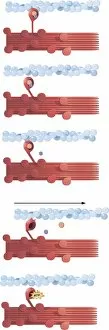
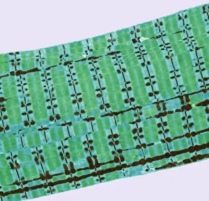
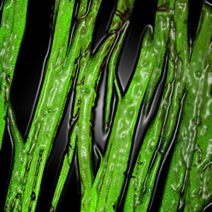
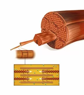
Myosin: The Mighty Molecular Motor of Muscle Contraction In the intricate world of muscle biology, myosin takes center stage as a vital player in the fascinating process of muscle contraction. This confocal light micrograph showcases the awe-inspiring beauty within heart muscle, highlighting its crucial role in sustaining life. Delving deeper into muscle fiber structure, an artwork captures the complexity and elegance of this microscopic marvel. Myosin molecules intricately interlock with actin filaments to generate force and enable movement, creating a symphony of motion within our bodies. Illustrations vividly depict how myosin powers muscle contraction. With labels guiding us through each step, we witness the coordinated dance between these molecular motor proteins and actin filaments that results in muscular movement - from flexing our biceps to pumping blood through our veins. However, sometimes even mighty they can face challenges. A close-up reveals details of deep back muscles experiencing sprain, strain, or spasm – reminders that proper care is essential for maintaining their health and functionality. Zooming further into the microscopic realm unveils torn muscle fibers surrounded by healing stages. It serves as a testament to both the resilience and regenerative capacity possessed by our body's remarkable machinery. Examining individual components brings us face-to-face with myosin itself – F006/9618 molecule stands tall as a molecular motor protein responsible for powering countless contractions throughout our lifetime. Its counterparts include Myosin molecule F006/9255 and Myosin fragment molecule F006/9245 - all working tirelessly behind-the-scenes to keep us moving effortlessly. An artistic representation called Actin Myosin Muscle Model (artwork C014/2661) offers insight into how these proteins interact on a larger scale. Their harmonious collaboration ensures smooth functioning across various muscles in our body while enabling strength and agility.