Home > Science > SEM
Coloured SEM of mitochondria & ER in a liver cell

![]()
Wall Art and Photo Gifts from Science Photo Library
Coloured SEM of mitochondria & ER in a liver cell
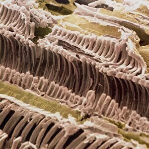
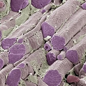
Mitochondria. Coloured Scanning Electron Micro- graph (SEM) of mitochondria and rough endoplasmic reticulum in a liver cell. The internal structure of a hepatocyte or liver cell is crowded with organelles, due to the metabolic activities of the cell. Mitochondria (pink) are sectioned through to show folds or cristae; they are sites of cell respiration and store energy. Rough endoplasmic reticulum (green) forms pathways in the cell; it is studded with ribosomes (dots, sites of protein synthesis). Liver cells detoxify waste products in blood, store glycogen and synthesize proteins. Magnification: x28, 000 at 6x7cm size. x36, 000 at 4x5ins
Science Photo Library features Science and Medical images including photos and illustrations
Media ID 6401099
© PROFESSOR P.M. MOTTA, G. MACCHIARELLI, S.A NOTTOLA/SCIENCE PHOTO LIBRARY
Cell Structure Cytology Endoplasmic Reticulum Hepatocyte Liver Cell Mitochondria Rough Rough Endoplasmic Reticulum Micro Biology
EDITORS COMMENTS
This print showcases the intricate internal structure of a liver cell, specifically highlighting the mitochondria and rough endoplasmic reticulum. The hepatocyte, or liver cell, is bustling with organelles due to its metabolic activities. In this image, the mitochondria are beautifully sectioned through to reveal their folds or cristae. These pink structures serve as crucial sites for cell respiration and energy storage. The green-colored rough endoplasmic reticulum forms pathways within the cell and is adorned with ribosomes (dots), which play a vital role in protein synthesis. Liver cells have multifaceted functions such as detoxifying waste products from the blood, storing glycogen, and synthesizing proteins. At a magnification of x28,000 for a 6x7cm size print or x36,000 for a 4x5ins print, this colored scanning electron micrograph (SEM) provides an extraordinary level of detail. It offers viewers an opportunity to appreciate the complexity and beauty found within our own bodies at microscopic levels. This remarkable image belongs to Science Photo Library's collection that focuses on various aspects of biology including cytology and microbiology. It serves as a testament to both scientific exploration and artistic appreciation by capturing the awe-inspiring intricacies hidden within our cells.
MADE IN THE UK
Safe Shipping with 30 Day Money Back Guarantee
FREE PERSONALISATION*
We are proud to offer a range of customisation features including Personalised Captions, Color Filters and Picture Zoom Tools
SECURE PAYMENTS
We happily accept a wide range of payment options so you can pay for the things you need in the way that is most convenient for you
* Options may vary by product and licensing agreement. Zoomed Pictures can be adjusted in the Basket.