Fraud Blocker Skip to main content

Connective Tissue Collection

Connective tissue is a remarkable network that holds our bodies together, providing support and structure to various organs and systems

Background imageConnective Tissue Collection: Anatomy of human knee joint

Anatomy of human knee joint

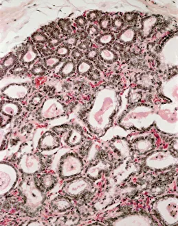
Background imageConnective Tissue Collection: Lactating breast tissue, light micrograph

Lactating breast tissue, light micrograph
Lactating breast tissue. Light micrograph of a section through healthy breast (mammary gland) tissue during lactation. Glandular tissue in the breast has proliferated to produce spaces (alveoli)

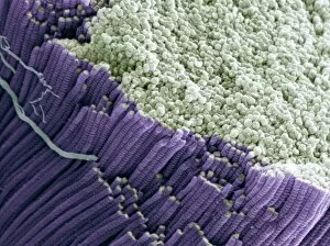
Background imageConnective Tissue Collection: Tendon, SEM

Tendon, SEM
Tendon, coloured scanning electron micrograph (SEM), showing bundles of collagen fibres. Magnification: x5000 when printed at 10 centimetres wide

Background imageConnective Tissue Collection: Outer ankle ligaments, artwork C013 / 4452

Outer ankle ligaments, artwork C013 / 4452
Outer ankle ligaments. Computer artwork of the bones and ligaments (white) of the right foot and ankle seen from the side

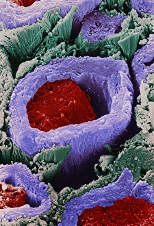
Background imageConnective Tissue Collection: Coloured SEM of a section through nerve fibres

Coloured SEM of a section through nerve fibres
Nerve fibres. Coloured scanning electron micrograph (SEM) of a section through bundles of nerve fibres (red). Each is within a sheath, or perineurium (blue)

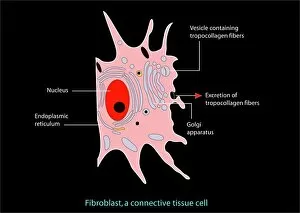
Background imageConnective Tissue Collection: Fibroblast cell, artwork

Fibroblast cell, artwork
Fibroblast cell. Computer artwork of a fibroblast excreting collagen fibres (tropocollagen). Fibroblasts are cells that produce connective tissue such as collagen (tropocollagen)

Background imageConnective Tissue Collection: Collagen fibres

Collagen fibres. Coloured transmission electron micrograph (TEM) of a section through healthy collagen fibres from human skin

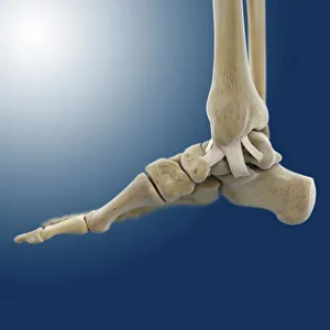
Background imageConnective Tissue Collection: Inner ankle ligaments, artwork C013 / 4451

Inner ankle ligaments, artwork C013 / 4451
Inner ankle ligaments. Computer artwork of the bones and ligaments (white) of the right foot and ankle seen from the side

Background imageConnective Tissue Collection: Red blood cells, computer artwork

Red blood cells, computer artwork
Red blood cells. Computer artwork of human red blood cells (erythrocytes) in a blood vessel. Red blood cells are biconcave, giving them a large surface area for gas exchange, and highly elastic

Background imageConnective Tissue Collection: Human tooth anatomy, artwork

Human tooth anatomy, artwork
Human tooth anatomy. Artwork of a cross-section through a human tooth to show its anatomical structure. The two main areas are the crown (above the gum-line) and the root (embedded in the gum)

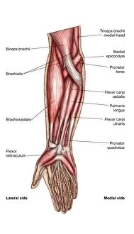
Background imageConnective Tissue Collection: Anatomy of human forearm muscles, superficial anterior view

Anatomy of human forearm muscles, superficial anterior view

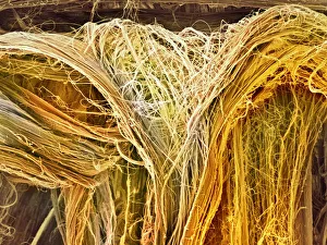
Background imageConnective Tissue Collection: Scanning electron microscope (SEM) of tendon

Scanning electron microscope (SEM) of tendon
Anatomy, Biology, Close-Up, Color Image, Connective Tissue, Fiber, Ful, SEM, 85757662

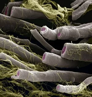
Background imageConnective Tissue Collection: Myelinated nerve fibers

Myelinated nerve fibers
Axon, Backgrounds, Biology, Color Image, Connective Tissue, Endoneuri, SEM, 85758245

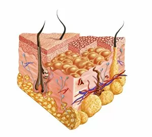
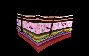
Background imageConnective Tissue Collection: Anatomy of the human skin

Anatomy of the human skin

Background imageConnective Tissue Collection: Anatomy of the female breast

Anatomy of the female breast

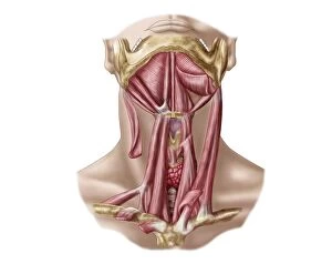
Background imageConnective Tissue Collection: Anatomy of human hyoid bone and muscles, anterior view

Anatomy of human hyoid bone and muscles, anterior view

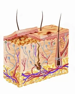
Background imageConnective Tissue Collection: Diagram showing anatomy of human skin

Diagram showing anatomy of human skin

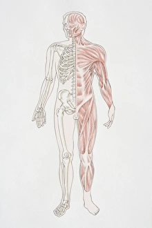
Background imageConnective Tissue Collection: Diagram illustrating the human musculo-skeletal system

Diagram illustrating the human musculo-skeletal system

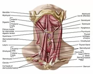
Background imageConnective Tissue Collection: Anatomy of human hyoid bone and neck muscles, anterior view

Anatomy of human hyoid bone and neck muscles, anterior view

Background imageConnective Tissue Collection: Retina with macular degeneration

Retina with macular degeneration

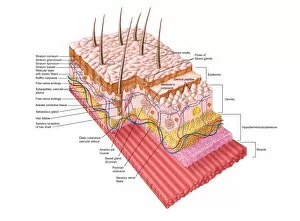
Background imageConnective Tissue Collection: Detailed cutaway diagram of human skin

Detailed cutaway diagram of human skin

Background imageConnective Tissue Collection: Anatomy of forearm muscles, anterior view, middle

Anatomy of forearm muscles, anterior view, middle

Background imageConnective Tissue Collection: Anatomy of human forearm muscles, deep anterior view

Anatomy of human forearm muscles, deep anterior view

Background imageConnective Tissue Collection: Different types of acne, non-inflammatory and inflammatory

Different types of acne, non-inflammatory and inflammatory

Background imageConnective Tissue Collection: Anatomy of the structure and layers of the stomach wall

Anatomy of the structure and layers of the stomach wall

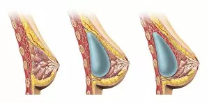
Background imageConnective Tissue Collection: Woman breast implant cross section

Woman breast implant cross section. Comparison diagram, under and over pectoral muscle

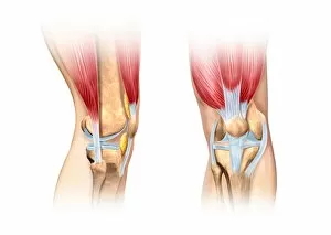
Background imageConnective Tissue Collection: Human knee cutaway illustration

Human knee cutaway illustration
Cutaway illustration of human knee showing detailed side and front views

Background imageConnective Tissue Collection: Conceptual image of the layers of human skin

Conceptual image of the layers of human skin

Background imageConnective Tissue Collection: Fascia of the lower leg, 1831 artwork

Fascia of the lower leg, 1831 artwork
Fascia of the lower leg, anterior (left) and posterior (right) views. Fascia are fibrous connective tissues that surround muscles and other tissues

Background imageConnective Tissue Collection: Trachea lining, SEM C015 / 9936

Trachea lining, SEM C015 / 9936
Trachea lining. Coloured scanning electron micrograph (SEM) of a section through the lining of the trachea (wind pipe). The trachea links the larynx (voice box) to the lungs

Background imageConnective Tissue Collection: Cartilage cell, TEM C014 / 1433

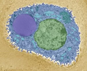
Cartilage cell, TEM C014 / 1433
Cartilage cell. Transmission electron micrograph (TEM) of a section through a chondrocyte cell from hyaline cartilage of the trachea (windpipe)

Background imageConnective Tissue Collection: Cartilage cell, TEM C014 / 1434

Cartilage cell, TEM C014 / 1434
Cartilage cell. Transmission electron micrograph (TEM) of a section through a chondrocyte cell from hyaline cartilage of the trachea (windpipe)

Background imageConnective Tissue Collection: Arm anatomy, artwork C013 / 4583

Arm anatomy, artwork C013 / 4583
Arm anatomy. Computer artwork of an oblique anterior view looking up a human arm, showing muscles and tendons (brown and white), bones and ligaments (white), arteries and veins (red and blue)

Background imageConnective Tissue Collection: Buttock muscles, artwork C013 / 4414

Buttock muscles, artwork C013 / 4414
Buttock muscles. Computer artwork of a posterior view of the hips showing some of the muscles (red) of the buttocks. These are shown attached to the femurs (thigh bones) and the pelvis bones

Background imageConnective Tissue Collection: Bladder epithelium, light micrograph

Bladder epithelium, light micrograph
Bladder epithelium. Light micrograph of a vertical section through the wall of the urinary bladder. The inner surface is at top

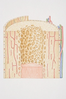
Background imageConnective Tissue Collection: Cross-section diagram of a human long bone

Cross-section diagram of a human long bone

Background imageConnective Tissue Collection: Heart with arteries showing cholesterol in one and plaque in the other

Heart with arteries showing cholesterol in one and plaque in the other
Heart with arteries showing cholesterol in one artery and atherosclerotic plaque in the other

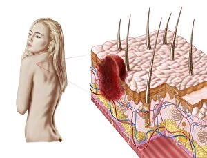
Background imageConnective Tissue Collection: Illustration of an atypical growth on the skin that could be a sign of skin cancer

Illustration of an atypical growth on the skin that could be a sign of skin cancer
Illustration of an atypical growth on the skin that could be a warning sign of skin cancer. Sudden change in the appearance, shape, color, size

Background imageConnective Tissue Collection: Detail of human knee showing insertion of arthroscopic instruments

Detail of human knee showing insertion of arthroscopic instruments

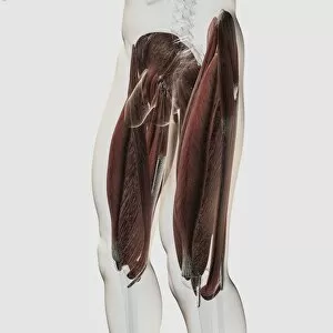
Background imageConnective Tissue Collection: Male muscle anatomy of the human legs, side view

Male muscle anatomy of the human legs, side view

Background imageConnective Tissue Collection: Arteries on heart showing atherosclerotic plaque in an artery

Arteries on heart showing atherosclerotic plaque in an artery

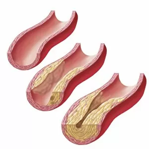
Background imageConnective Tissue Collection: Build-up of atherosclerotic plaque in an artery

Build-up of atherosclerotic plaque in an artery

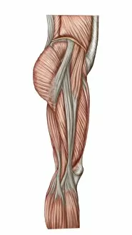
Background imageConnective Tissue Collection: Anatomy of human thigh muscles, anterior view

Anatomy of human thigh muscles, anterior view

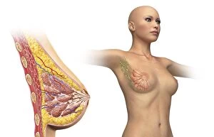
Background imageConnective Tissue Collection: Cutaway view of female breast with woman figure showing lymphatic glands

Cutaway view of female breast with woman figure showing lymphatic glands
Woman breast cutaway, cross section diagram with woman figure showing lymphatic glands

Background imageConnective Tissue Collection: Acute coronary syndrome - microvascular obstruction

Acute coronary syndrome - microvascular obstruction

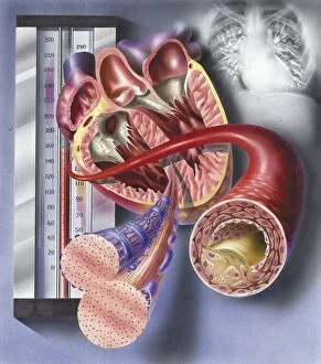
Background imageConnective Tissue Collection: Interior view of heart with detail of muscle cells and atherosclerotic artery

Interior view of heart with detail of muscle cells and atherosclerotic artery. Chest x-ray in background

Background imageConnective Tissue Collection: Retina of a normal eye

Retina of a normal eye

Background imageConnective Tissue Collection: Thrombus forming on valve within vein

Thrombus forming on valve within vein


All Professionally Made to Order for Quick Shipping

Connective tissue is a remarkable network that holds our bodies together, providing support and structure to various organs and systems. In the anatomy of the human knee joint, connective tissue plays a crucial role in maintaining stability and allowing smooth movement. Lactating breast tissue, as seen under a light microscope, showcases the intricate arrangement of connective fibers that aid in milk production. Examining tendons through scanning electron microscopy reveals their strong composition primarily made up of collagen fibers. These tough yet flexible strands provide resilience and enable efficient transmission of forces between muscles and bones. Artwork depicting outer ankle ligaments (C013 / 4452) illustrates how connective tissue safeguards joints from excessive movements while ensuring proper alignment during physical activities. Similarly, inner ankle ligaments (C013 / 4451) contribute to joint stability by connecting bones within the ankle region. Even on a microscopic level, connective tissue continues to amaze us. Computer artwork showcasing red blood cells highlights their vital role in transporting oxygen throughout our body via an intricate network of capillaries embedded within this specialized type of connective tissue. Delving into human tooth anatomy through artwork unveils another aspect where they are present – supporting structures like periodontal ligament that anchor teeth firmly within the jawbone. Fibroblast cells depicted in artwork demonstrate their pivotal function in synthesizing extracellular matrix components such as collagen and elastin – essential for wound healing and maintaining overall tissue integrity. Mesenchymal stem cells captured using scanning electron microscopy exhibit immense potential for regenerative medicine due to their ability to differentiate into various cell types found within different types of connective tissues. The optic nerve fibers imaged under SEM highlight how delicate yet resilient these structures are, responsible for transmitting visual information from our eyes to the brain with utmost precision thanks to surrounding supportive connective tissues. Lastly, examining fat tissue at high magnification reveals its unique structure composed mainly of adipocytes, which store energy and provide insulation.