Fraud Blocker Skip to main content

False Coloured Collection

"Revealing the Unseen: Exploring the World Through False Colors" Step back in time to medieval alchemy

Background imageFalse Coloured Collection: E. coli bacteria, SEM

E. coli bacteria, SEM
E. coli bacteria. Coloured scanning electron micrograph (SEM) of Escherichia coli bacteria (purple) taken from the small intestine of a child. E

Background imageFalse Coloured Collection: Helicobacter pylori bacteria

Helicobacter pylori bacteria
False-colour transmission electron micrograph of the bacterium Helicobacter pylori (formerly called Campylobacter pyloridis)

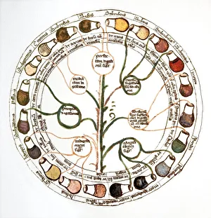
Background imageFalse Coloured Collection: Medieval urine wheel

Medieval urine wheel. Labelled in Latin, this 15th century diagram shows some of the possible colours of urine (outer edge of circle) to help doctors diagnose urine-related diseases

Background imageFalse Coloured Collection: Orion nebula

Orion nebula. Coloured composite infrared and visible light image of the Orion nebula M42. This emission nebula, a cloud of gas and dust in which starbirth takes place

Background imageFalse Coloured Collection: Particle tracks

Particle tracks
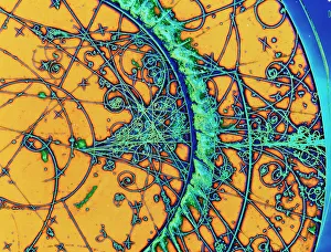
Subatomic particle tracks. Coloured bubble chamber photograph showing tracks left by subatomic particles from a particle accelerator at CERN, the European particle physics laboratory at Geneva

Background imageFalse Coloured Collection: X and Y chromosomes

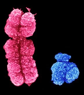
X and Y chromosomes, coloured scanning electron micrograph (SEM). Humans have 46 chromosomes in total: 23 inherited from the mother and 23 from the father

Background imageFalse Coloured Collection: Horses skull

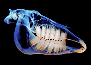
Horses skull, coloured X-ray

Background imageFalse Coloured Collection: Nerve cell, SEM

Nerve cell, SEM
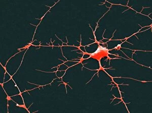
Nerve cell. Coloured scanning electron micrograph (SEM) of a nerve cell (neuron). Neurons are responsible for passing information around the central nervous system (CNS)

Background imageFalse Coloured Collection: Cat tongue surface, SEM

Cat tongue surface, SEM
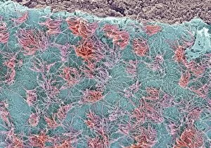
Cats tongue. Coloured scanning electron micrograph (SEM) of the surface of a cats (family Felidae) tongue. The tongue is covered in many backward facing papillae (projections)

Background imageFalse Coloured Collection: Martian landscape, Spirit rover image

Martian landscape, Spirit rover image
Martian landscape. False-colour image of the Martian surface taken by NASAs Mars exploration rover Spirit between the 23rd and 24th November 2005. Part of the rover can be seen at bottom centre

Background imageFalse Coloured Collection: Uterus lining during menstruation, SEM

Uterus lining during menstruation, SEM
Uterus during menstruation. Coloured scanning electron micrograph (SEM) of the lining of the uterus being shed during menstruation

Background imageFalse Coloured Collection: Brain anatomy, MRI scan

Brain anatomy, MRI scan
Brain anatomy. Coloured magnetic resonance imaging (MRI) scan of the human head from the side. The sagittal scan has halved the brain, revealing the internal anatomy

Background imageFalse Coloured Collection: Salmonella bacteria, SEM

Salmonella bacteria, SEM
Salmonella bacteria, coloured scanning electron micrograph (SEM). These Gram-negative rod-shaped bacteria can cause food poisoning when eaten in contaminated food

Background imageFalse Coloured Collection: Vascular bundle, SEM

Vascular bundle, SEM
Vascular bundle. Coloured scanning electron micrograph (SEM) of a section through a vascular bundle from a rootlet of a dicotyledon plant

Background imageFalse Coloured Collection: Geranium anther, SEM

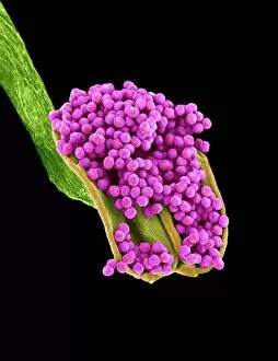
Geranium anther, SEM
Geranium anther. Coloured scanning electron micrograph (SEM) of pollen (pink) on the anther of a geranium flower (Geranium sp.)

Background imageFalse Coloured Collection: Dahlia flower pollen, SEM

Dahlia flower pollen, SEM
Dahlia flower pollen. Coloured scanning electron micrograph (SEM) of a pollen grain (orange) from a Dahlia cultorum flower. A pollen grain contains a male sex cell from a flowering plant

Background imageFalse Coloured Collection: Tardigrade, SEM

Tardigrade, SEM
Water bear. Coloured scanning electron micrograph (SEM) of a water bear, or tardigrade (phylum Tardigrada). Water bears are small, water-dwelling

Background imageFalse Coloured Collection: Head louse, SEM

Head louse, SEM
Head louse. Coloured scanning electron micrograph (SEM) of a head louse (Pediculus humanus capitis) clinging to a human hair. Head lice measure 2-3 millimetres in length

Background imageFalse Coloured Collection: Acrosphaera radiolarian, SEM

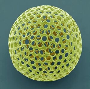
Acrosphaera radiolarian, SEM
Acrosphaera radiolarian. Coloured scanning electron micrograph (SEM) of the shell of a Acrosphaera sp. radiolarian. Radiolaria are single-celled protozoans that are found in marine plankton

Background imageFalse Coloured Collection: Dog tapeworm head, SEM

Dog tapeworm head, SEM
Dog tapeworm head. Coloured scanning electron micrograph (SEM) of the head (scolex) of a dog tapeworm (Taenia pisiformis)

Background imageFalse Coloured Collection: Ant, SEM

Ant, SEM
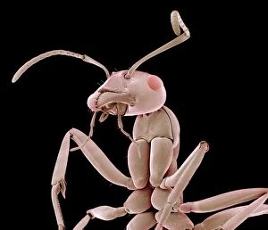
Ant. Coloured scanning electron micrograph (SEM) of an ant (family Formicidae). One of its compound eyes (red) is seen, as well as two long antennae that are mounted on the head between the eyes

Background imageFalse Coloured Collection: Red-barbed ant, SEM

Red-barbed ant, SEM
Red-barbed ant (Formica rufibarbis), coloured scanning electron micrograph (SEM). This ant is common in mainland Europe, but rare in the UK

Background imageFalse Coloured Collection: Small intestine, SEM

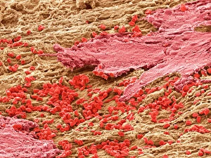
Small intestine, SEM
Small intestine. Coloured scanning electron micrograph (SEM) of a biopsy from the small intestines of a child. The small intestine runs from the stomach to the large intestine

Background imageFalse Coloured Collection: Synapse nerve junction, TEM

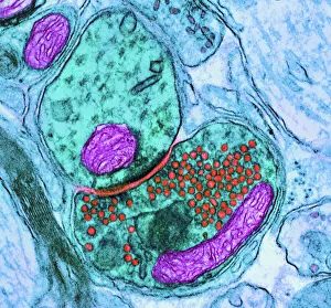
Synapse nerve junction, TEM
Synapse. Coloured transmission electron micrograph (TEM) of a synapse, a junction between two nerve cells, in the brain. At a synapse an electrical signal is transmitted from one cell to the next in

Background imageFalse Coloured Collection: Seahorse skeleton

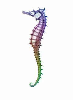
Seahorse skeleton. Coloured x-ray of a seahorse (Hippocampus sp.). Seahorse populations are under threat due to the fishing industry

Background imageFalse Coloured Collection: Knee joint prosthesis, X-ray

Knee joint prosthesis, X-ray
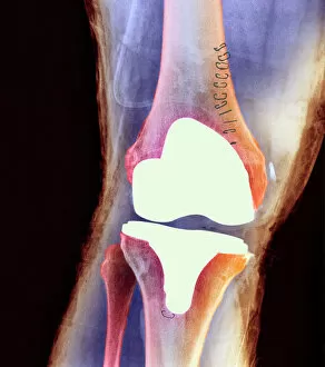
Knee joint replacement. Coloured X-ray of the knee of a 56 year old man (front view) showing an artificial (prosthetic) joint replacing the knee joint

Background imageFalse Coloured Collection: Foot of a chameleon

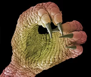
Foot of a chameleon. Coloured scanning electron micrograph (SEM) of the foot of a chameleon. Chameleons are the only lizards with zygodactyle feet, or with toes in a pincer arrangement

Background imageFalse Coloured Collection: Neck vertebrae extended, X-ray

Neck vertebrae extended, X-ray
Bending of the neck. Coloured X-ray of a side view of the neck of a man showing extension of healthy cervical vertebrae (bones)

Background imageFalse Coloured Collection: Woven fabric, SEM

Woven fabric, SEM
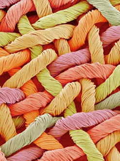
Woven fabric fibres. Coloured scanning electron micrograph (SEM) of synthetic fibres woven to form clothes. Nylon and polyester are common synthetic clothing materials

Background imageFalse Coloured Collection: Rough endoplasmic reticulum, TEM

Rough endoplasmic reticulum, TEM
Rough endoplasmic reticulum, coloured transmission electron micrograph (TEM). This section shows the rough endoplasmic reticulum (ER, folds, centre), a membranous structure that occurs in cells

Background imageFalse Coloured Collection: Calcareous phytoplankton, SEM

Calcareous phytoplankton, SEM
Calcareous phytoplankton. Coloured scanning electron micrograph (SEM) of the external mineralised structures (coccospheres) of small marine algal organisms called coccolithophores

Background imageFalse Coloured Collection: Total hip replacement, X-ray

Total hip replacement, X-ray
Total hip replacement. Coloured frontal X-ray of a female pelvis with a total hip replacement (white, lower right). The replacement hip consists of a ball and shaft

Background imageFalse Coloured Collection: Normal skull, X-ray

Normal skull, X-ray
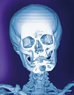
Normal skull, coloured frontal 3D computed tomography (CT) scan

Background imageFalse Coloured Collection: Fractured ankle, X-ray

Fractured ankle, X-ray
Fractured ankle. Coloured profile X-ray of a distal fibula fracture (upper centre). The fibula is the smaller leg bone running down from top centre. The larger bone is the tibia

Background imageFalse Coloured Collection: Primate skulls

Primate skulls. Coloured x-rays of the skulls of a gorilla (Gorilla gorilla, left), chimpanzee (Pan trogoldytes, centre) and human (Homo sapien, left)

Background imageFalse Coloured Collection: Brain surface, SEM

Brain surface, SEM
Brain surface. Coloured scanning electron micrograph (SEM) of the surface of a brain ventricle. Ventricles are cavities within the brain that are continuous with the central canal of the spinal cord

Background imageFalse Coloured Collection: Pollen grains, SEM

Pollen grains, SEM
Pollen grains. Coloured scanning electron micrographs (SEM) of pollen grains from a variety of plants

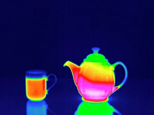
Background imageFalse Coloured Collection: Teapot and hot drink, thermogram

Teapot and hot drink, thermogram
Teapot and hot drink. Thermogram of a teapot containing a hot beverage, next to a hot drink. Thermography records surface temperatures by detecting the long-wavelength radiation emitted by an object

Background imageFalse Coloured Collection: Salmonella bacteria, SEM

Salmonella bacteria, SEM
Salmonella bacteria, coloured scanning electron micrograph (SEM). Each rod-shaped structure is an individual bacterium. These bacteria occur mainly in human and animal intestines

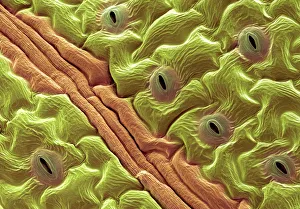
Background imageFalse Coloured Collection: Leaf pores, SEM

Leaf pores, SEM
Leaf pores. Coloured scanning electron micrograph (SEM) of stomata (holes) on the surface of a leaf. These structures perform a similar function to the pores in human skin

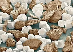
Background imageFalse Coloured Collection: Salt grains and ground peppercorn, SEM

Salt grains and ground peppercorn, SEM
Salt grains and ground peppercorn, coloured scanning electron micrograph (SEM). Each salt (sodium chloride) crystal (white) is composed of a cubic lattice of sodium and chloride ions

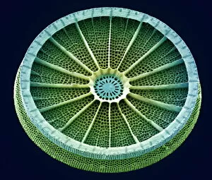
Background imageFalse Coloured Collection: Diatom, SEM

Diatom, SEM
Diatom. Coloured scanning electron micrograph (SEM) of the Arachnoidiscus sp. diatom. The diatoms are a group of photosynthetic, single- celled algae containing about 10, 000 species

Background imageFalse Coloured Collection: Diatom, SEM

Diatom, SEM
Diatom. Coloured scanning electron micrograph (SEM) of a Stephanopyxis sp. diatom. Diatoms are single-celled photosynthetic algae, of which there are about 100, 000 species

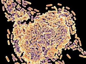
Background imageFalse Coloured Collection: E. coli bacteria, SEM

E. coli bacteria, SEM
E. coli bacteria. Coloured scanning electron micrograph (SEM) of Escherichia coli bacteria. These bacteria are a normal part of the intestinal flora in humans and other animals

Background imageFalse Coloured Collection: Diatom, SEM

Diatom, SEM
Diatom. Coloured scanning electron micrograph (SEM) of a Triceratium morlandii diatom. Diatoms are single-celled photosynthetic algae, of which there are about 100, 000 species

Background imageFalse Coloured Collection: Culex mosquito, SEM

Culex mosquito, SEM
Culex mosquito. Coloured scanning electron micrograph (SEM) of a Culex sp. mosquito. The mosquitos head is dominated by its large compound eyes (brown spheres)

Background imageFalse Coloured Collection: Waxing gibbous Moon, computer-enhanced

Waxing gibbous Moon, computer-enhanced image. The colour saturation of this image has been enhanced to accentuate the natural variation in colour of the Moons surface

Background imageFalse Coloured Collection: 19th-century tin mine, Cornwall

19th-century tin mine, Cornwall. Artwork of miners and walkways at the Botallack Copper and Tin Mine at St Just, Cornwall, England. This mine, under various names, dates back to 1721

Background imageFalse Coloured Collection: Triggerfish skeleton, X-ray

Triggerfish skeleton, X-ray
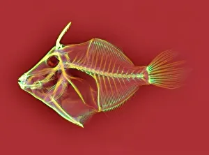
Triggerfish skeleton. Coloured X-ray of a triggerfish (family Balistidae). Its name originates from its ability to erect the first two dorsal spines (upper left), which can then lock or unlock

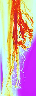
Background imageFalse Coloured Collection: Varicose veins, X-ray

Varicose veins, X-ray
Varicose veins. Coloured X-ray of varicose veins in the calf of a fifty-five-year-old male. The veins (red), which carry blood back to the heart, are swollen, irregular and distorted

Background imageFalse Coloured Collection: Embryonic stem cell and needle, SEM

Embryonic stem cell and needle, SEM
Embryonic stem cell and needle. Coloured scanning electron micrograph (SEM) of an embryonic stem cell (ESC) sitting in the eye of a needle

Background imageFalse Coloured Collection: Candida fungus, SEM

Candida fungus, SEM
Candida fungus. Coloured scanning electron micrograph (SEM) of budding threads (hyphae) of a Candida fungus. The budding areas are where asexual reproduction is producing new fungus cells

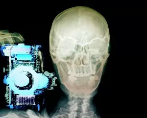
Background imageFalse Coloured Collection: Person with a camera, X-ray

Person with a camera, X-ray
Person with a camera. Coloured, frontal X-ray of a person holding a camera (blue, left). The persons skull (centre) and neck bones (vertebrae, lower centre) can be seen

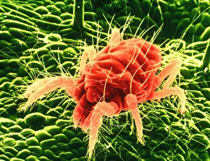
Background imageFalse Coloured Collection: False-colour SEM of Tetranychus, a red spider mite

False-colour SEM of Tetranychus, a red spider mite

Background imageFalse Coloured Collection: Sheep tick, SEM

Sheep tick, SEM
Sheep tick. Coloured scanning electron micrograph (SEM) of a sheep tick (Ixodes ricinus). The ticks mouthparts are between its two front legs

Background imageFalse Coloured Collection: Regenerating nerve cell, TEM

Regenerating nerve cell, TEM
Regenerating nerve cell. Coloured transmission electron micrograph (TEM) of a section through a nerve axon (blue) regenerating within a Schwann cell (light brown)

Background imageFalse Coloured Collection: Myelination of nerve fibres, TEM

Myelination of nerve fibres, TEM
Myelination of nerve fibres. Coloured transmission electron micrograph (TEM) of Schwann cells (blue, with brown nuclei) insulating nerve fibres (axons, pink) with a myelin sheath

Background imageFalse Coloured Collection: Water bear, SEM

Water bear, SEM
Water bear. Coloured scanning electron micrograph (SEM) of a water bear (Macrobiotus sp.), or tardigrade. This tiny invertebrate lives in aquatic and semi-aquatic habitats such as lichen

Background imageFalse Coloured Collection: Gallbladder surface, SEM

Gallbladder surface, SEM
Gallbladder. Coloured scanning electron micrograph (SEM) of the surface of the gallbladder showing the numerous microvilli that cover the columnar epithelium

Background imageFalse Coloured Collection: Moth proboscis, SEM

Moth proboscis, SEM
Moth proboscis. Coloured scanning electron micrograph (SEM) of the coiled proboscis of a moth (order Lepidoptera). The proboscis is an elongated part of the mouth

Background imageFalse Coloured Collection: Honey bee, SEM

Honey bee, SEM
Honey bee (Apis mellifera), coloured scanning electron micrograph (SEM). The head and one of the two compound eyes is at lower left

Background imageFalse Coloured Collection: Head of a honey bee, SEM

Head of a honey bee, SEM
Head of a honey bee (Apis mellifera), coloured scanning electron micrograph (SEM). The compound eyes (one seen) are either side of the antennae

Background imageFalse Coloured Collection: Norovirus particles, TEM

Norovirus particles, TEM
Norovirus particles. Coloured transmission electron micrograph (TEM) of norovirus particles. Norovirus is a genus of RNA (ribonucleic acid)

Background imageFalse Coloured Collection: Pseudomonas aeruginosa bacteria, SEM

Pseudomonas aeruginosa bacteria, SEM
Pseudomonas aeruginosa bacteria, coloured scanning electron micrograph (SEM). These Gram-negative rod-shaped bacteria are found in soil and water, and as normal flora in the human intestine

Background imageFalse Coloured Collection: Dividing yeast cells, SEM

Dividing yeast cells, SEM
Dividing yeast cells. Coloured scanning electron micrograph (SEM) of Schizosaccharomyces pombe yeast cells dividing. S. pombe is a single-celled fungus that is studied widely as a model organism for

Background imageFalse Coloured Collection: Dividing yeast cells, SEM

Dividing yeast cells, SEM
Dividing yeast cells. Coloured scanning electron micrograph (SEM) of Schizosaccharomyces pombe yeast cells dividing. S. pombe is a single-celled fungus that is studied widely as a model organism for

Background imageFalse Coloured Collection: Bacteria infecting a macrophage, SEM

Bacteria infecting a macrophage, SEM
Bacteria infecting a macrophage. Coloured scanning electron micrograph (SEM) of Mycobacterium tuberculosis bacteria (purple) infecting a macrophage white blood cell

Background imageFalse Coloured Collection: Pseudomonas aeruginosa bacteria, SEM

Pseudomonas aeruginosa bacteria, SEM
Pseudomonas aeruginosa bacteria, coloured scanning electron micrograph (SEM). These Gram-negative rod-shaped bacteria are found in soil and water, and as normal flora in the human intestine

Background imageFalse Coloured Collection: E. coli bacterium, TEM

E. coli bacterium, TEM
E. coli bacterium, coloured transmission electron micrograph (TEM). E. coli are Gram-negative rod-shaped bacteria that are part of the normal flora of the human gut

Background imageFalse Coloured Collection: Fat cells, TEM

Fat cells, TEM
Fat cells. Coloured transmission electron micrograph (TEM) of a section through fat (adipose) tissue, consisting of fat-storing cells (adipocytes, green) supported by connective tissue

Background imageFalse Coloured Collection: Basophil white blood cell

Basophil white blood cell. Coloured transmission electron micrograph (TEM) of a basophil white blood cell. Basophils are the smallest and rarest of the white blood cells

Background imageFalse Coloured Collection: False-colour photo of total solar eclipse, 1979

False-colour photo of total solar eclipse, 1979
False-colour photograph of the total solar eclipse of February 26, 1979. The colour has been added by a photographic technique called isophote mapping

Background imageFalse Coloured Collection: Regenerating nerve cell, TEM

Regenerating nerve cell, TEM
Regenerating nerve cell. Coloured transmission electron micrograph (TEM) of a section through a nerve axon (yellow) regenerating within a Schwann cell (blue). The Schwann cells nucleus is black

Background imageFalse Coloured Collection: Stem cells, SEM

Stem cells, SEM
Stem cells, coloured scanning electron micrograph (SEM). Stem cells can differentiate into any other cell type. There are three main types of mammalian stem cell: embryonic stem cells

Background imageFalse Coloured Collection: Motor neurons, light micrograph

Motor neurons, light micrograph. Motor neurons are responsible for passing information around the central nervous system (CNS) and from the CNS to the rest of the body

Background imageFalse Coloured Collection: Geranium pollen, SEM

Geranium pollen, SEM
Geranium pollen. Coloured scanning electron micrograph (SEM) of pollen (pink) on the stamen of a geranium flower (Geranium sp.)

Background imageFalse Coloured Collection: Bacteria found on mobile phone, SEM

Bacteria found on mobile phone, SEM
Bacteria found on mobile phone. Coloured scanning electron micrograph (SEM) of bacteria cultured from a mobile phone. Tests have revealed the average handset carries 18 times more potentially harmful

Background imageFalse Coloured Collection: Gregor Mendel, Austrian botanist

Gregor Mendel, Austrian botanist
Gregor Johann Mendel (1822-1884), Austrian botanist and founder of genetics. Mendel, the abbot of an abbey in Brno, carried out breeding experiments with pea plants (held in hand)

Background imageFalse Coloured Collection: Dermestid beetle, SEM

Dermestid beetle, SEM
Dermestid beetle. Scanning electron micrograph (SEM(=) of a dermestid beetle. Dermestidae are a family of Coleoptera that are commonly referred to as skin beetles

Background imageFalse Coloured Collection: Needle playing a record, SEM

Needle playing a record, SEM
Needle playing a record. Coloured scanning electron micrograph (SEM) of the needle (stylus) of a record player in a groove on a record. A record is used to store sound

Background imageFalse Coloured Collection: 19th-century gold mining, Australia

19th-century gold mining, Australia. Artwork of Chinese workers excavating and washing gold ores at a gold mine in Australia

Background imageFalse Coloured Collection: Medical zodiac, 15th century diagram

Medical zodiac, 15th century diagram
Medical zodiac. 15th century diagram with Latin text illustrating how the human body relates to the zodiac signs. Such information was used in medical practices, such as bloodletting

Background imageFalse Coloured Collection: Galileo and his daughter Maria Celeste

Galileo and his daughter Maria Celeste
Galilei Galileo (1564-1642), Italian physicist and astronomer, being guided by his daughter Maria Celeste (1600-1634), a nun

Background imageFalse Coloured Collection: Normal spine, X-ray

Normal spine, X-ray
Normal spine. Coloured profile X-ray showing bones of the spine (vertebrae, centre). The front of the body is at right. The ribs (upper right) and the pelvis (bottom) are also seen


All Professionally Made to Order for Quick Shipping

"Revealing the Unseen: Exploring the World Through False Colors" Step back in time to medieval alchemy, where a peculiar contraption known as the "Medieval urine wheel" was used to extract hidden secrets from bodily fluids. Fast forward to modern times and witness how science has evolved, unveiling breathtaking wonders like the Orion Nebula. Delve into the microscopic realm, where particle tracks paint a mesmerizing picture of subatomic interactions. Journey across Martian landscapes through the eyes of Spirit rover images, capturing otherworldly vistas that ignite our imagination. Peering into our own genetic blueprint, false colors bring clarity to X and Y chromosomes, unraveling mysteries within our very cells. A horse's skull takes on an ethereal glow as we explore its intricate structure using advanced imaging techniques. Witness synapse nerve junctions come alive with vibrant hues under a transmission electron microscope (TEM), revealing connections that shape our thoughts and actions. A person holding a camera becomes an enigma when captured through X-ray technology – their inner world exposed for all to see. Marvel at tardigrades' resilience as they navigate their microscopic universe under scanning electron microscopy (SEM). Calcareous phytoplankton dance with color in intricate patterns while nerve cells reveal their complexity in stunning detail. Travel back in time to 19th-century Cornwall and discover the harsh reality of tin mining brought vividly to life through false colors – reminding us of humanity's enduring spirit even amidst adversity. In this captivating journey through various realms – from ancient alchemy to distant galaxies, minuscule particles to majestic landscapes – false colors unlock hidden dimensions beyond what meets the eye, inviting us all into a world brimming with wonder and discovery.

© Copyright Media Storehouse, All Rights Reserved 2003 - 2024
Facebook     Pinterest     Card Payments     Reviews IO