
Myofibrils Collection
Myofibrils: The Powerhouses of Muscle Contraction From the intricate eye muscles to the mighty heart muscle


All Professionally Made to Order for Quick Shipping
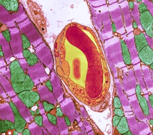
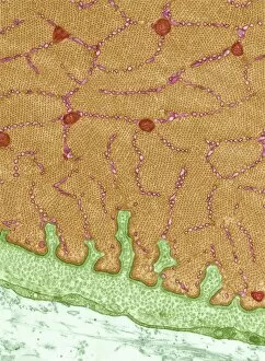
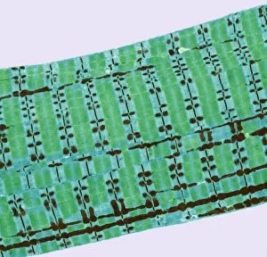
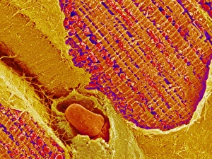
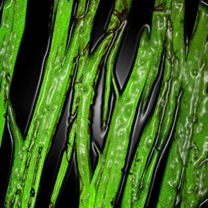
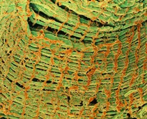
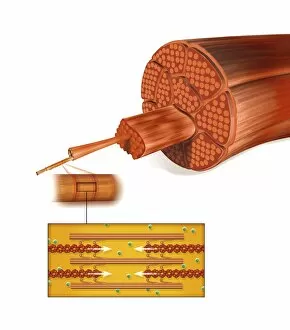
Myofibrils: The Powerhouses of Muscle Contraction From the intricate eye muscles to the mighty heart muscle, myofibrils play a crucial role in our body's movement and function. These microscopic structures are responsible for generating force and enabling us to perform various activities. In the eye muscle, as seen through TEM C014 / 1468, myofibrils form a complex network that allows precise control over our vision. Similarly, in the confocal light micrograph of the heart muscle, we witness how myofibrils contribute to its rhythmic contractions. The artwork depicting muscle fiber structure showcases the organized arrangement of these contractile units within skeletal and cardiac muscles. Through TEM images like those of cardiac muscle and capillary or skeletal muscle (TEM), we gain insight into their detailed architecture at a cellular level. But what fuels these incredible machines? Diagrams illustrating sugar uptake in muscles reveal how glucose is transported into myofibrils to provide energy for contraction. This vital process ensures smooth functioning during physical activity. Artwork such as Actin Myosin Muscle Model (C014 / 2661) helps visualize how actin and myosin filaments interact within myofibrils to generate force. Their coordinated movements create muscular contractions necessary for everyday tasks. Another fascinating aspect is revealed through artwork showcasing neuromuscular junctions - where nerves meet muscles. This connection enables communication between neurons and myofibrils, allowing precise control over movement. As we delve deeper into understanding these remarkable structures, TEM images like Eye Muscle (C014 / 1467) and Eye Muscle (C014 / 1466) showcase their unique features specific to ocular functions. Whether it be powering our eyesight or ensuring our hearts beat steadily, myofibrils prove themselves indispensable time after time. With each new discovery aided by advanced imaging techniques like TEM or artistic representations highlighting their complexity, we continue to unravel the mysteries of these microscopic powerhouses.