
Framed Print > Science > SEM
Framed Print : Small intestine villus, SEM


![]()
Framed Photos from Science Photo Library
Small intestine villus, SEM
Small intestine villus. Coloured scanning electron micrograph (SEM) of a freeze fracture section through a villus from the mucosal lining of the small intestine. Villi are finger-like projections that increase the surface area of a structure. Microvilli, just visible across upper centre, further increase the surface area available for food absorption. The outer surface of a villus is mostly columnar epithelium (red). It contains numerous goblet cells (dark pink), which secrete mucus to lubricate food & prevent self-digestion. Within the goblet cells individual mucin granules are seen. Magnification: x2000 at 6x7cm size
Science Photo Library features Science and Medical images including photos and illustrations
Media ID 6450661
© STEVE GSCHMEISSNER/SCIENCE PHOTO LIBRARY
Absorption Absorptive Alimentary Canal Columnar Digestion Digestive System Enterocyte Enterocytes Epithelial Epithelium Fractured Goblet Cell Histology Intestinal Lining Membrane Microvilli Mucosa Mucous Mucus Secretory Small Intestine Surface Tissue Villi Villus Cells
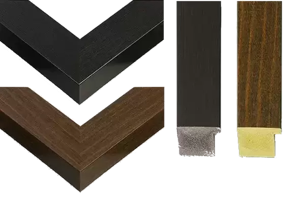
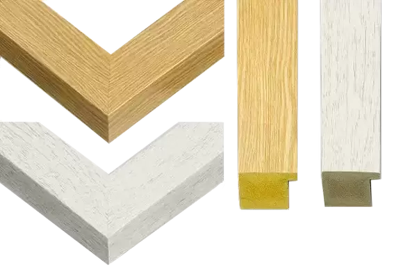
14"x12" (38x32cm) Modern Frame
Discover the intricacies of the human body with our Media Storehouse Framed Prints featuring the captivating "Small Intestine Villus, SEM" image from Science Photo Library. This coloured scanning electron micrograph (SEM) reveals the complex structure of a villus from the mucosal lining of the small intestine in stunning detail. Bring the wonders of science into your home or office with our high-quality, frameless prints, meticulously crafted to preserve the vibrant colours and sharp details of this remarkable image. Each print comes with a certificate of authenticity and is ready to hang, making it an excellent addition to any space dedicated to science, education, or artistic exploration.
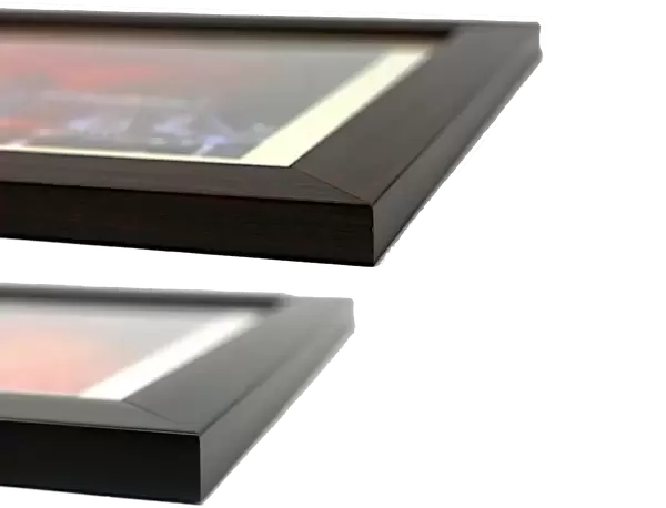
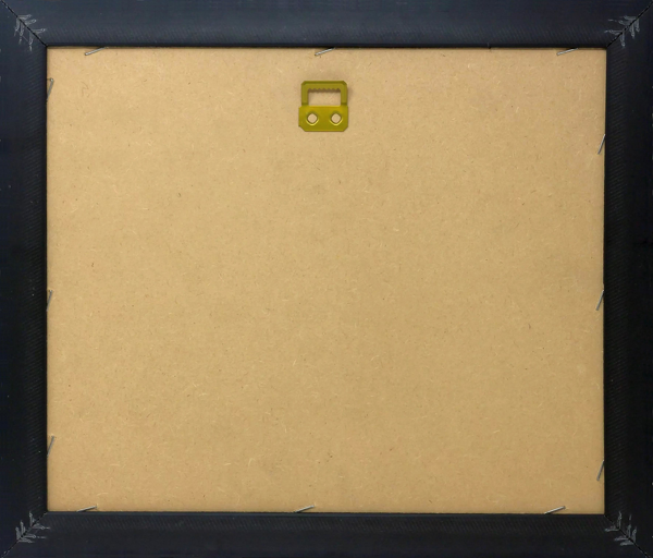
Wood effect frame, card mounted, 10x8 archival quality photo print. Overall outside dimensions 14x12 inches (38x32cm). Environmentally and ozone friendly, 40mm wide x 15mm Polycore® moulding has the look of real wood, is durable and light and easy to hang. Biodegradable and made with non-chlorinated gases (no toxic fumes) it is efficient; producing 100 tons of polystyrene can save 300 tons of trees! Prints are glazed with lightweight, shatterproof, optical clarity acrylic (providing the same general protection from the environment as glass). The back is stapled hardboard with a sawtooth hanger attached. Note: To minimise original artwork cropping, for optimum layout, and to ensure print is secure, the visible print may be marginally smaller
Contemporary Framed and Mounted Prints - Professionally Made and Ready to Hang
Estimated Image Size (if not cropped) is 24.4cm x 19.6cm (9.6" x 7.7")
Estimated Product Size is 37.6cm x 32.5cm (14.8" x 12.8")
These are individually made so all sizes are approximate
Artwork printed orientated as per the preview above, with landscape (horizontal) orientation to match the source image.
EDITORS COMMENTS
This print showcases the intricate details of a small intestine villus, captured through a scanning electron microscope (SEM). The image reveals the vibrant colors and textures of this vital organ's mucosal lining. Villi, which resemble finger-like projections, can be seen protruding from the surface, significantly increasing its overall surface area for efficient food absorption. Additionally, microvilli are faintly visible across the upper center of the image, further enhancing this absorptive capacity. The outer layer of the villus primarily consists of columnar epithelium in a striking red hue. This layer contains numerous goblet cells that secrete mucus to lubricate food and prevent self-digestion. Within these goblet cells, individual mucin granules can be observed under magnification. With a magnification level of x2000 at 6x7cm size, this SEM image provides an up-close look at healthy tissue within our digestive system. It offers valuable insights into the histology and anatomy of our gut while highlighting key components such as membranes, epithelial cells (enterocytes), and secretory functions. This remarkable photograph is part of Science Photo Library's extensive collection on human body anatomy and serves as a testament to their commitment to providing high-quality scientific imagery for educational purposes.
MADE IN THE UK
Safe Shipping with 30 Day Money Back Guarantee
FREE PERSONALISATION*
We are proud to offer a range of customisation features including Personalised Captions, Color Filters and Picture Zoom Tools
SECURE PAYMENTS
We happily accept a wide range of payment options so you can pay for the things you need in the way that is most convenient for you
* Options may vary by product and licensing agreement. Zoomed Pictures can be adjusted in the Basket.