
Absorptive Collection
"Absorptive Marvels: Unveiling the Intricate World of Absorption" Step into a mesmerizing journey as we explore the fascinating realm of absorption


All Professionally Made to Order for Quick Shipping
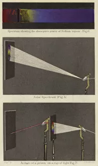
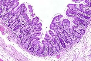
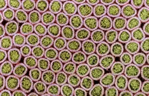
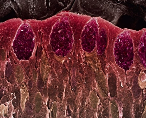
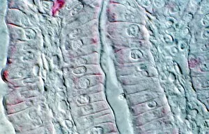
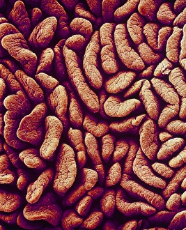
"Absorptive Marvels: Unveiling the Intricate World of Absorption" Step into a mesmerizing journey as we explore the fascinating realm of absorption, where Solar Spectrum (colour litho) unveils its captivating hues. Delve deep into the microscopic wonders that make our bodies efficient absorbers. Intestinal villi, SEM - Like delicate fingers reaching out for nutrients, these tiny projections line our intestines and play a crucial role in absorbing vital substances. Behold the intricate beauty of Foetal skin, SEM C016 / 9045. Witness how even at such an early stage, our skin possesses absorbent qualities that protect and nourish us. Intestinal microvilli, SEM C016 / 9066 & C016 / 9067 - These hair-like structures cover the surface of intestinal cells like a dense forest. Their purpose? To maximize absorption by increasing surface area and ensuring no nutrient goes unnoticed. Marvel at Foetal skin's resilience under SEM C016 / 9044. This close-up reveals its unique ability to absorb essential elements while providing protection to developing life within. Colon, light micrograph C016 / 0516 - A glimpse into this organ's inner workings showcases its remarkable capacity for water absorption. Witness nature's efficiency in action. Prepare to be amazed by Intestinal microvilli under SEM C014 / 1452 & C014 / 1451. These high-resolution images capture their elegant structure as they tirelessly work to extract every ounce of nutrition from our food. Peer through the lens of TEM at Intestinal microvilli C014/1454 &C014/1453 – revealing their ultra-fine details with astonishing clarity. Discover how these minute structures facilitate nutrient transport on a cellular level. Finally, marvel at the grandeur of Intestinal lining under SEM – an awe-inspiring tapestry woven with precision and designed for optimal absorption. Every fold tells a story of the body's remarkable ability to extract life-sustaining elements.