
Absorption Collection
"Absorption: Unveiling the Mysteries of Nature's Intricate Processes" Delving into the intricate world of absorption


All Professionally Made to Order for Quick Shipping
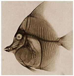
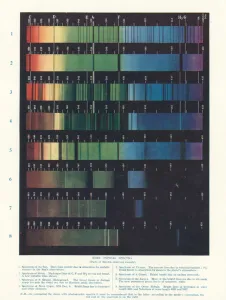
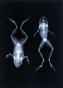
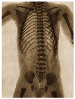
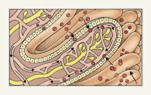
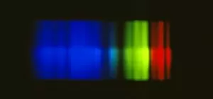
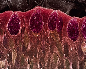
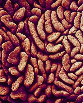
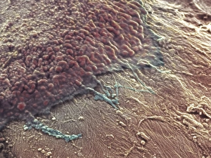
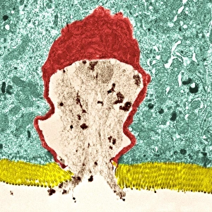
"Absorption: Unveiling the Mysteries of Nature's Intricate Processes" Delving into the intricate world of absorption, we encounter a mesmerizing illustration showcasing intestinal villi, where nutrients are absorbed and transported to nourish our bodies. The Antonov An-26SM 52+06 aircraft takes center stage as it demonstrates its ability to absorb cargo effortlessly, highlighting how absorption plays a crucial role in transportation. A captivating X-ray from c. 1890 reveals the inner structure of a fish, shedding light on how organisms absorb essential minerals from their environment for growth and survival. Exploring beyond our earthly realm, visible emission spectra of astronomical objects captivate us with their vibrant colors, reminding us that even celestial bodies undergo processes of absorption. St Francis in Ecstasy (c. 1615-18) by an unknown artist depicts spiritual absorption as the saint transcends worldly concerns and immerses himself in divine ecstasy. Pioneering scientist Joseph Maria Eder's early X-ray photo of frogs (1896) showcases his groundbreaking work on radiography, revealing how this technology revolutionized medical diagnosis by absorbing images within living organisms. Journeying back to c. 1890 through an X-ray lens captures moments frozen in time - a child's hand, an ankle fracture, and a fractured wrist - illustrating both trauma and healing processes involving bone tissue absorption. Peering inside the human torso through another vintage X-ray image from c. 1890 unravels secrets hidden beneath our skin; organs working harmoniously while absorbing vital substances for bodily functions. The Pictorial Museum of Animated Nature enchants us with its engraved pages depicting diverse creatures absorbing sustenance from their surroundings – nature's delicate balance at play. The Crab Nebula dazzles astronomers with its radiant beauty; formed from remnants of a supernova explosion centuries ago—a cosmic absorption and transformation that continues to captivate our imaginations.