
Villus Collection
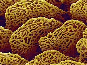
The small intestine is a fascinating organ in the human digestive system, and one of its key components are the villi


All Professionally Made to Order for Quick Shipping
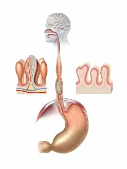
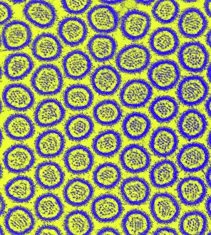
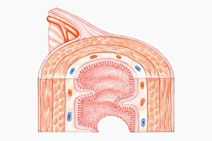
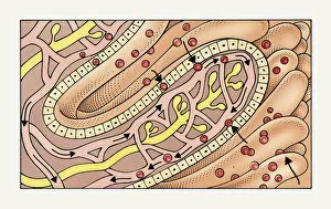
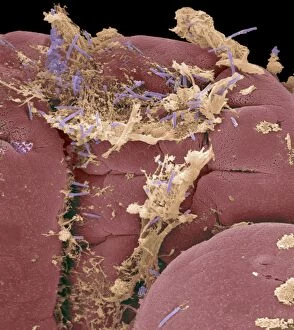
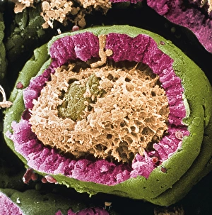
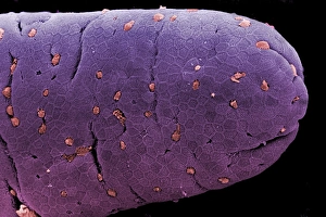
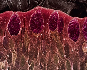
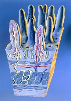
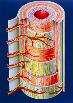
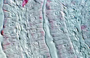
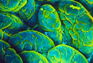
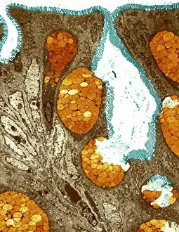
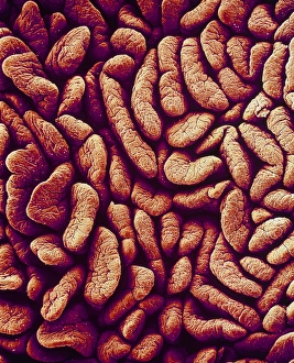
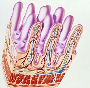
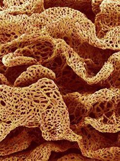
The small intestine is a fascinating organ in the human digestive system, and one of its key components are the villi. These tiny finger-like projections play a crucial role in absorbing nutrients from our food. When observed under a scanning electron microscope (SEM), the intricate structure of these villi becomes apparent. They line the inner surface of the intestinal wall, forming an elaborate network that increases the surface area for absorption. This artwork depicting the human digestive system showcases their importance beautifully. Zooming in further with a transmission electron microscope (TEM), we can see another level of detail - microvilli covering each individual villus. These microscopic structures greatly enhance nutrient absorption by further increasing surface area. In this SEM image, we get an up-close look at intestinal villi themselves. Their unique shape and arrangement contribute to efficient digestion and absorption processes within our bodies. A cross-section illustration provides us with insight into how these villi fit into the overall structure of the small intestine. We can see layers including muscle tissue, plicae (folds), and prominently displayed are those vital villi responsible for nutrient uptake. Another captivating illustration demonstrates how peptides and amino acids are absorbed into capillaries through these remarkable structures known as villi. It's incredible to think about all that happens on such a microscopic scale. Even arteries have not escaped their influence. A microscopic view reveals how intestinal villi extend inside blood vessels, ensuring efficient transportation of absorbed nutrients throughout our body systems. Conceptual images help us visualize just how important these little protrusions truly are. They represent life-sustaining functions occurring within our bodies every day – extracting essential substances from food to nourish ourselves. Microscopic views provide yet another glimpse into this world within us; they reveal intricate details hidden beneath what we perceive externally. The delicate nature of these intestinal villi is awe-inspiring when seen up close like this.