
Acrylic Blox > Science > SEM
Acrylic Blox : Small intestine villus, SEM


![]()
Mounted Prints from Science Photo Library
Small intestine villus, SEM
Small intestine villus. Coloured scanning electron micrograph (SEM) of a freeze fracture section through a villus from the mucosal lining of the small intestine. Villi are finger-like projections that increase the surface area of a structure. Microvilli, just visible across upper centre, further increase the surface area available for food absorption. The outer surface of a villus is mostly columnar epithelium (red). It contains numerous goblet cells (dark pink), which secrete mucus to lubricate food & prevent self-digestion. Within the goblet cells individual mucin granules are seen. Magnification: x2000 at 6x7cm size
Science Photo Library features Science and Medical images including photos and illustrations
Media ID 6450661
© STEVE GSCHMEISSNER/SCIENCE PHOTO LIBRARY
Absorption Absorptive Alimentary Canal Columnar Digestion Digestive System Enterocyte Enterocytes Epithelial Epithelium Fractured Goblet Cell Histology Intestinal Lining Membrane Microvilli Mucosa Mucous Mucus Secretory Small Intestine Surface Tissue Villi Villus Cells
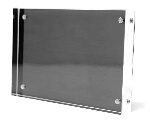
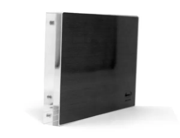
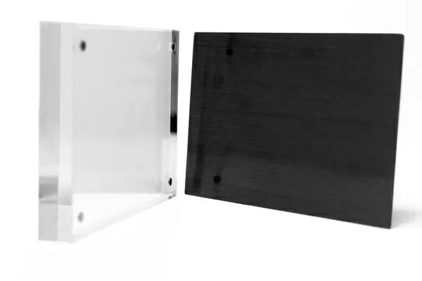
8"x6" (20x15cm) Acrylic Blox
Your photographic print is held in place by magnets and a micro thin sheet of metal covering the back of a 20mm piece of clear acrylic. Your print is held in place with magnets so can easily be replaced if needed.
Streamlined, one sided modern and attractive table top print
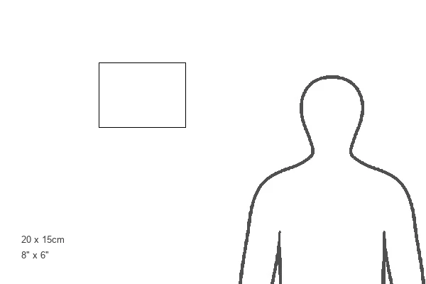
Estimated Product Size is 20.3cm x 15.2cm (8" x 6")
These are individually made so all sizes are approximate
Artwork printed orientated as per the preview above, with landscape (horizontal) orientation to match the source image.
EDITORS COMMENTS
This print showcases the intricate details of a small intestine villus, captured through a scanning electron microscope (SEM). The image reveals the vibrant colors and textures of this vital organ's mucosal lining. Villi, which resemble finger-like projections, can be seen protruding from the surface, significantly increasing its overall surface area for efficient food absorption. Additionally, microvilli are faintly visible across the upper center of the image, further enhancing this absorptive capacity. The outer layer of the villus primarily consists of columnar epithelium in a striking red hue. This layer contains numerous goblet cells that secrete mucus to lubricate food and prevent self-digestion. Within these goblet cells, individual mucin granules can be observed under magnification. With a magnification level of x2000 at 6x7cm size, this SEM image provides an up-close look at healthy tissue within our digestive system. It offers valuable insights into the histology and anatomy of our gut while highlighting key components such as membranes, epithelial cells (enterocytes), and secretory functions. This remarkable photograph is part of Science Photo Library's extensive collection on human body anatomy and serves as a testament to their commitment to providing high-quality scientific imagery for educational purposes.
MADE IN THE UK
Safe Shipping with 30 Day Money Back Guarantee
FREE PERSONALISATION*
We are proud to offer a range of customisation features including Personalised Captions, Color Filters and Picture Zoom Tools
SECURE PAYMENTS
We happily accept a wide range of payment options so you can pay for the things you need in the way that is most convenient for you
* Options may vary by product and licensing agreement. Zoomed Pictures can be adjusted in the Basket.