Fraud Blocker Skip to main content

Cells Collection

"Exploring the Intricacies of Cells: From Cerebellum Tissue to Geranium Anther" Delving into the complexity of cells

Background imageCells Collection: Cerebellum tissue, light micrograph

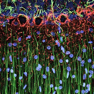
Cerebellum tissue, light micrograph
Cerebellum tissue. Confocal light micrograph of a section through the cerebellum of the brain. Purkinje cells, a type of neuron (nerve cell), are red

Background imageCells Collection: Hippocampus brain tissue

Hippocampus brain tissue
Hippocampus tissue. Light micrograph of a sagittal (side view) section through the hippocampus of the brain showing the nerve cells within it

Background imageCells Collection: Nerve and glial cells, light micrograph

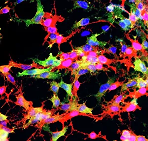
Nerve and glial cells, light micrograph
Nerve and glial cells, fluorescence light micrograph. These are neural stem cells that have differentiated into neurons (nerve cells, blue) and glial cells (support cells, red)

Background imageCells Collection: A prison guard on patrol in B wing of Britains top security prison Parkhurst

A prison guard on patrol in B wing of Britains top security prison Parkhurst on the Isle of Wight November 1984

Background imageCells Collection: Nerve cells

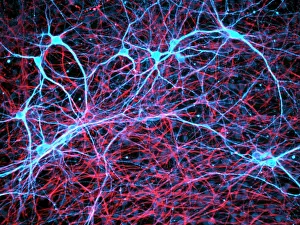
Nerve cells
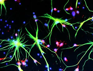
Immunofluorescent Light Micrograph of neuron cells and astrocytes in mammalian spinal cord. Here, neuron cells stain red: the cell body appears pink

Background imageCells Collection: Pentonville Prison, Islington, North London

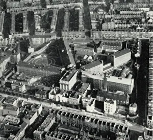
Pentonville Prison, Islington, North London
An aerial view of Pentonville Prison in Islington, North London. The prison was opened in 1842 as the new Model Prison, on the separate system

Background imageCells Collection: Wandsworth Prison, south west London

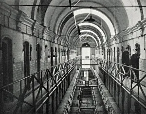
Wandsworth Prison, south west London
A galleried wing of Wandsworth Prison in south west London. Two warders inspect the cells

Background imageCells Collection: Cut-away Diagram of the Tower of London, 1913

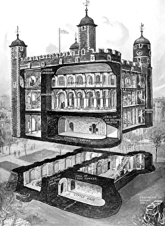
Cut-away Diagram of the Tower of London, 1913
Cut-away illustration showing a number of the rooms of the Tower of London, including the Chapel of St. John, the Armouries and the cells of Guy Fawkes, Sir Thomas More and Sir Walter Raleigh

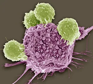
Background imageCells Collection: T lymphocytes and cancer cell, SEM C001 / 1679

T lymphocytes and cancer cell, SEM C001 / 1679
T lymphocytes and cancer cell. Coloured scanning electron micrograph (SEM) of T lymphocyte cells (green) attached to a cancer cell

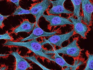
Background imageCells Collection: HeLa cells, light micrograph C017 / 8299

HeLa cells, light micrograph C017 / 8299
HeLa cells, multiphoton fluorescence micrograph (MFM). The cell nuclei, which contain the cells genetic information, are purple. Microtubules are blue and actin microfilaments are red

Background imageCells Collection: Aylesbury Prison 1900

Aylesbury Prison 1900
Aylesbury Prison corridor B3 1900 Date: 1900

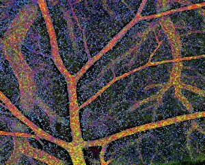
Background imageCells Collection: Brain tissue blood supply

Brain tissue blood supply. Light micrograph of a section through cortex tissue from a brain, showing the blood vessels (branching) that supply it

Background imageCells Collection: Dicotyledon plant stem, light micrograph

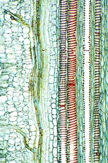
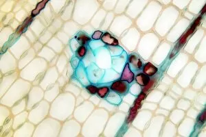
Dicotyledon plant stem, light micrograph
Dicotyledon plant stem. Light micrograph of a longitudinal radial section through a typical dicotyledon stem. The section has passed through a vascular bundle and other stem tissue

Background imageCells Collection: Geranium anther, SEM

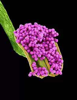
Geranium anther, SEM
Geranium anther. Coloured scanning electron micrograph (SEM) of pollen (pink) on the anther of a geranium flower (Geranium sp.)

Background imageCells Collection: Botanik Digitalis purpurea L. Fingerhut 160: 1

Botanik Digitalis purpurea L. Fingerhut 160: 1
Foxglove leaf. Coloured scanning electron micrograph (SEM) of a section through a leaf from a foxglove (Digitalis purpurea) plant, showing its hairs (trichomes)

Background imageCells Collection: Aylesbury Prison 1900

Aylesbury Prison 1900
Aylesbury Prison cell 1900 Date: 1900

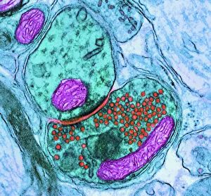
Background imageCells Collection: Synapse nerve junction, TEM

Synapse nerve junction, TEM
Synapse. Coloured transmission electron micrograph (TEM) of a synapse, a junction between two nerve cells, in the brain. At a synapse an electrical signal is transmitted from one cell to the next in

Background imageCells Collection: Lime tree stem, light micrograph

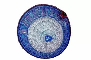
Lime tree stem, light micrograph
Lime tree stem. Light micrograph of a section through the stem of a lime tree (Tilia europaea). The outer epidermis has been shed and replaced by a layer of cork (dark red)

Background imageCells Collection: Glial stem cell culture, light micrograph

Glial stem cell culture, light micrograph
Glial stem cell culture. Fluorescent light micrograph of glial stem cells producing the protein NG2 (red) as they mature. These stem cells can differentiate into several types of glial cells

Background imageCells Collection: Plant cell

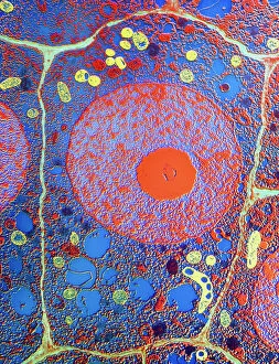
Plant cell
False-colour transmission electron micrograph (TEM) of a cell in the root tip of a maize plant, Zea mays. Plant cells are distinct from animal cells in having an additional external envelope

Background imageCells Collection: State Penitentiary at Stateville, Joliet, Illinois, USA

State Penitentiary at Stateville, Joliet, Illinois, USA
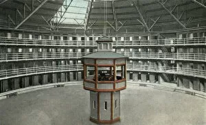
Interior view of a cell block at the Illinois State Penitentiary at Stateville, near Joliet, Illinois, USA. The prison was designed using the panopticon principle promoted by Jeremy Bentham

Background imageCells Collection: Cerebral cortex nerve cells

Cerebral cortex nerve cells. Confocal light micrograph of neurons (nerve cells, red) and glial cells (support cells, gold) from the cerebral cortex

Background imageCells Collection: Electricity / Voltaic Pile

Electricity / Voltaic Pile
VOLTAIC PILE The first workable device for generating a consistent flow of electricity, invented in 1799 by Alessandro Volta

Background imageCells Collection: Cell structure

Cell structure. Confocal light micrograph of cultured endothelial cells. A fluorescent dye has been used to show the cell structure

Background imageCells Collection: Embryonic stem cell and needle, SEM

Embryonic stem cell and needle, SEM
Embryonic stem cell and needle. Coloured scanning electron micrograph (SEM) of an embryonic stem cell (ESC) sitting in the eye of a needle

Background imageCells Collection: HeLa cells, light micrograph C017 / 8298

HeLa cells, light micrograph C017 / 8298
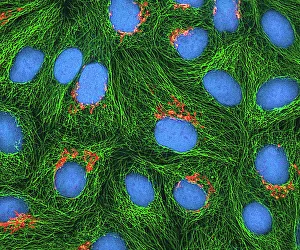
HeLa cells, multiphoton fluorescence micrograph (MFM). The cell nuclei, which contain the cells genetic information, are blue. Golgi bodies, which modify and package proteins, are orange

Background imageCells Collection: Dendritic cells, artwork

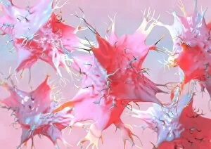
Dendritic cells, artwork. Dendritic cells, a type of white blood cell, are called antigen presenting cells (APCs) and are found in the bodys tissues

Background imageCells Collection: TEM of human macrophage

TEM of human macrophage

Background imageCells Collection: Blood cells

Blood cells, computer artwork. Red blood cells (red), or erythrocytes, are biconcave disc-shaped cells that are responsible for supplying tissues with oxygen

Background imageCells Collection: Pine pollen grains, light micrograph

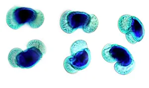
Pine pollen grains, light micrograph
Pine pollen grains. Light micrograph of a section through winged pollen grains (microspores) from a pine (Pinus sp.) tree

Background imageCells Collection: Pine stem, light micrograph

Pine stem, light micrograph
Pine stem. Light micrograph of a section through the stem of a pine (Pinus sp.) tree, showing xylem tissue. The xylem is made up of tracheid cells (light pink)

Background imageCells Collection: Athlete physiology, artwork

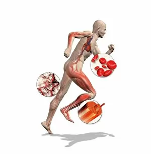
Athlete physiology, artwork
Athlete physiology. Computer artwork of a male athlete, with enlargements to show the important physiological enhancements typical of most athletes

Background imageCells Collection: Cell types, artwork

Cell types, artwork
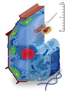
Cell types. Cutaway artwork with a scale bar (upper right, in micrometres) showing the relative sizes of eukaryotic cells (those containing a nucleus) and prokaryotic cells (those lacking a nucleus)

Background imageCells Collection: Neural stem cell culture

Neural stem cell culture. Fluorescent light micrograph of a group of neural stem cells (neurosphere) in culture. Neural stem cells are able to differentiate into neurons (nerve cells)

Background imageCells Collection: Dividing yeast cells, SEM

Dividing yeast cells, SEM
Dividing yeast cells. Coloured scanning electron micrograph (SEM) of Schizosaccharomyces pombe yeast cells dividing. S. pombe is a single-celled fungus that is studied widely as a model organism for

Background imageCells Collection: Dividing yeast cells, SEM

Dividing yeast cells, SEM
Dividing yeast cells. Coloured scanning electron micrograph (SEM) of Schizosaccharomyces pombe yeast cells dividing. S. pombe is a single-celled fungus that is studied widely as a model organism for

Background imageCells Collection: Bacteria infecting a macrophage, SEM

Bacteria infecting a macrophage, SEM
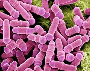
Bacteria infecting a macrophage. Coloured scanning electron micrograph (SEM) of Mycobacterium tuberculosis bacteria (purple) infecting a macrophage white blood cell

Background imageCells Collection: Pseudomonas aeruginosa bacteria, SEM

Pseudomonas aeruginosa bacteria, SEM
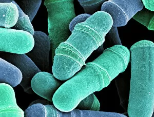
Pseudomonas aeruginosa bacteria, coloured scanning electron micrograph (SEM). These Gram-negative rod-shaped bacteria are found in soil and water, and as normal flora in the human intestine

Background imageCells Collection: Cerebellum tissue, light micrograph

Cerebellum tissue, light micrograph
Cerebellum tissue. Confocal light micrograph of a section through the cerebellum of the brain showing two types of glial cells (support cells); astrocytes (star-shaped)

Background imageCells Collection: Glial cells, confocal light micrograph

Glial cells, confocal light micrograph
Glial cells. Confocal light micrograph of glial cells from the cerebellum of the brain. Glial cells are nervous system cells that provide structural support and protection for neurons (nerve cells)

Background imageCells Collection: Fat cells, TEM

Fat cells, TEM
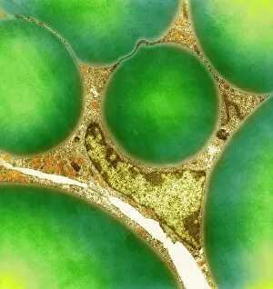
Fat cells. Coloured transmission electron micrograph (TEM) of a section through fat (adipose) tissue, consisting of fat-storing cells (adipocytes, green) supported by connective tissue

Background imageCells Collection: Coloured SEM of a white blood cell (lymphocyte)

Coloured SEM of a white blood cell (lymphocyte)
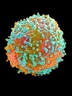
Lymphocyte blood cell. Coloured scanning electron micrograph (SEM) of a lymphocyte, a common type of white blood cell. Characteristic of this rounded cell are the long microvilli which project

Background imageCells Collection: Stem cells, SEM

Stem cells, SEM
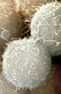
Stem cells, coloured scanning electron micrograph (SEM). Stem cells can differentiate into any other cell type. There are three main types of mammalian stem cell: embryonic stem cells

Background imageCells Collection: Motor neurons, light micrograph

Motor neurons, light micrograph. Motor neurons are responsible for passing information around the central nervous system (CNS) and from the CNS to the rest of the body

Background imageCells Collection: Geranium pollen, SEM

Geranium pollen, SEM
Geranium pollen. Coloured scanning electron micrograph (SEM) of pollen (pink) on the stamen of a geranium flower (Geranium sp.)

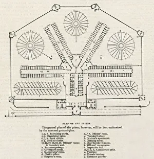
Background imageCells Collection: Plan Pentonville Prison

Plan Pentonville Prison
A plan of the floor layout of Pentonville, a model prison built in London in 1843, with corridors of cells radiating out from a central point & exercise yards

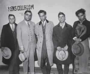
Background imageCells Collection: Joe Aiello (1891-1930) with his henchmen in police custody, 1928 (b / w photo)

Joe Aiello (1891-1930) with his henchmen in police custody, 1928 (b / w photo)
PNP252300 Joe Aiello (1891-1930) with his henchmen in police custody, 1928 (b/w photo) by American Photographer, (20th century); Private Collection; (add.info)

Background imageCells Collection: Solar panel with sun

Solar panel with sun

Background imageCells Collection: Osteocyte bone cell, SEM C016 / 9025

Osteocyte bone cell, SEM C016 / 9025
Osteocyte bone cell. Coloured scanning electron micrograph (SEM) of an osteocyte bone cell (blue) surrounded by bone tissue (pink)

Background imageCells Collection: HeLa cells, light micrograph C013 / 4774

HeLa cells, light micrograph C013 / 4774
HeLa cells. Multi-photon fluorescence light micrograph of a group of cultured HeLa cells, showing the cell nuclei, which contain the cells genetic information (DNA, red)

Background imageCells Collection: Human skin, illustration

Human skin, illustration
3d illustration of a cross section of a hair follicle with description

Background imageCells Collection: Picture No. 11675585

Picture No. 11675585
Scanning Electron micrograph (SEM)showing stomata on a Yew Leaf. Date:

Background imageCells Collection: Picture No. 11675528

Picture No. 11675528
Mouse Embryo at Eight Cell Stage. Date:

Background imageCells Collection: Picture No. 11675527

Picture No. 11675527
Mouse Embryo at Four Cell Stage & a Polar Body. Date:

Background imageCells Collection: Picture No. 11675526

Picture No. 11675526
Mouse Embryo at Two Cell Stage & a Polar Body. Date:

Background imageCells Collection: Picture No. 11675525

Picture No. 11675525
Mouse Embryo at Single Cell Stage and Polar Body. Date:

Background imageCells Collection: Picture No. 11675522

Picture No. 11675522
A human Ovum surrounded by Sperm - being fertilized. Date:

Background imageCells Collection: Picture No. 11675612

Picture No. 11675612
Scanning Electron Micrograph (SEM): Human White and Red Blood Cells. Date:

Background imageCells Collection: Picture No. 10876997

Picture No. 10876997
Scanning Electron Micrograph (SEM): Human Skin with Hair Follicles Date:

Background imageCells Collection: Dohle bodies in blood cell, micrograph

Dohle bodies in blood cell, micrograph
Dohle bodies in blood cell. Light micrograph of a neutrophil white blood cell (centre) with Dohle bodies. These are small inclusions within the cells cytoplasm

Background imageCells Collection: Blood coagulation cascade, artwork C016 / 9873

Blood coagulation cascade, artwork C016 / 9873
Blood coagulation cascade. Artwork of the biochemical cascade of blood chemicals and proteins during blood clotting (coagulation). The blood vessel and its layered wall is at upper left

Background imageCells Collection: English oak leaf pores, SEM

English oak leaf pores, SEM
English oak leaf pores. Coloured scanning electron micrograph (SEM) of stomata (round) on the underside of a leaf from an English oak (Quercus robur) tree

Background imageCells Collection: Pancreas anatomy, artwork

Pancreas anatomy, artwork
Pancreas anatomy. Cut-away computer artwork of the pancreas, which secretes digestive enzymes and the hormones insulin and glucagon, which regulate blood sugar levels

Background imageCells Collection: Maize root, light micrograph

Maize root, light micrograph
Maize root. Light micrograph of a section through the root of a maize plant (Zea mays) showing a vascular cylinder (centre)

Background imageCells Collection: Water lily leaf, light micrograph

Water lily leaf, light micrograph
Water lily leaf. Light micrograph of a transverse section through the leaf of a water lily (Nympha sp.) plant. All aquatic plants (hydrophytes) have a similar structure

Background imageCells Collection: Cell Block Interior, Wakefield Prison, West Yorkshire

Cell Block Interior, Wakefield Prison, West Yorkshire
The interior of a cell block at Wakefield Prison, West Yorkshire. Cells line the open galleries which have a safety net strung across to prevent inmates falling

Background imageCells Collection: Cerebellum tissue, light micrograph

Cerebellum tissue, light micrograph
Cerebellum tissue. Confocal light micrograph of a section through the cerebellum of the brain. Purkinje cells, a type of neuron (nerve cell), are red

Background imageCells Collection: IUD contraceptive and sperm cells

IUD contraceptive and sperm cells. Computer artwork of an intrauterine device (IUD, red and white, centre) in a womans uterus, surrounded by sperm cells (white, not to scale)

Background imageCells Collection: Antibodies attacking virus particles, illustration

Antibodies attacking virus particles, illustration
3d illustration of antibodies attacking virus particles in the bloodstream

Background imageCells Collection: Picture No. 11675582

Picture No. 11675582
Scanning Electron micrograph (SEM): Lily Pollen grains. Date:

Background imageCells Collection: Plasma cells, TEM

Plasma cells, TEM
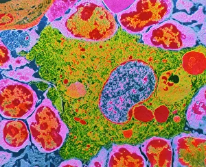
Plasma cells. Coloured transmission electron micrograph (TEM) of plasma cells. Plasma cells are mature B lymphocytes (white blood cells) that produce and secrete antibodies during an immune response

Background imageCells Collection: Nerve cells, SEM

Nerve cells, SEM
Nerve cells. Coloured scanning electron micrograph (SEM) of nerve cells, known as neurones. Nerve cells occur in the brain, spinal cord, and in ganglia

Background imageCells Collection: Picture No. 10873704

Picture No. 10873704
Sleeping Sickness Parasite in red blood cells (Trypanosoma sp. ) Date:

Background imageCells Collection: Acute promyelocytic leukaemia, micrograph

Acute promyelocytic leukaemia, micrograph
Acute promyelocytic leukaemia. Light micrograph of blood cells from bone marrow in a case of acute promyelocytic leukaemia. Leukaemia is a cancer where certain blood cells form in excess

Background imageCells Collection: Rod and cone cells of the eye, SEM C014 / 4866

Rod and cone cells of the eye, SEM C014 / 4866
Rod and cone cells of the eye. Coloured scanning electron micrograph (SEM) of rod and cone cells in the retina of a mammalian eye

Background imageCells Collection: Diatoms, SEM

Diatoms, SEM
Diatoms. Coloured scanning electron micrograph (SEM) of diatoms (Campylodiscus sp.). These microscopic, single-celled algae possess rigid and finely-sculpted cell walls known as frustules

Background imageCells Collection: Dividing cells

Dividing cells. Computer artwork of a spiral of cells during mitosis (nuclear division). During mitosis two daughter nuclei are formed from one parent nucleus

Background imageCells Collection: Pancreatic islet of Langerhans

Pancreatic islet of Langerhans
Islet of Langerhans. Light micrograph of a section through an islet of Langerhans (pale, centre) in pancreas tissue. This clump of secretory cells forms part of the endocrine system of the body

Background imageCells Collection: Red blood cells, SEM

Red blood cells, SEM
Red blood cells, coloured scanning electron micrograph (SEM). Red blood cells (erythrocytes) are biconcave, giving them a large surface area for gas exchange, and highly elastic

Background imageCells Collection: Astrocyte nerve cell

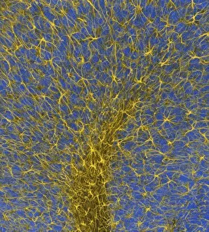
Astrocyte nerve cell. Fluorescent light micrograph of an astrocyte cell from a human brain. Intermediate filaments (IFs), part of the cells cytoskeleton, have been dyed green

Background imageCells Collection: Nerve cells, abstract artwork

Nerve cells, abstract artwork
Nerve cells. Abstract computer artwork of nerve cells, or neurons. Neurons are responsible for passing information around the central nervous system (CNS) and from the CNS to the rest of the body

Background imageCells Collection: Neutrophil cell trapping bacteria, SEM

Neutrophil cell trapping bacteria, SEM
Neutrophil cell trapping bacteria. Coloured scanning electron micrograph (SEM) of bacteria (rod-shaped) being trapped by a neutrophil cell

Background imageCells Collection: Kidney glomerulus, SEM

Kidney glomerulus, SEM
Kidney glomerulus, coloured scanning electron micrograph (SEM). Podocyte cells, which make up the surface of the glomerulus, are yellow. Magnification: x650 when printed at 10 centimetres wide

Background imageCells Collection: Lung tissue, TEM

Lung tissue, TEM
Lung tissue, coloured transmission electron micrograph (TEM). A single alveolus is at center. At top right is a pulmonary capillary containing a red blood cell (red) and a platelet (yellow)


All Professionally Made to Order for Quick Shipping

"Exploring the Intricacies of Cells: From Cerebellum Tissue to Geranium Anther" Delving into the complexity of cells, we examine cerebellum tissue through a captivating light micrograph. Behind the walls of Wandsworth Prison in southwest London, cells silently carry out their vital functions within inmates' bodies. Witnessing the intricate network of nerve and glial cells, a mesmerizing light micrograph reveals their interconnectedness. At the synapse nerve junction, captured by TEM imaging, we witness the fascinating communication between cells that allows our body to function seamlessly. Within Pentonville Prison in Islington, North London, hidden stories unfold as various cell types coexist amidst confinement. Shedding light on groundbreaking research, T lymphocytes and cancer they can magnified under SEM imaging - offering hope for future treatments (SEM C001 / 1679). Peering into hippocampus brain tissue unveils a world where countless cells work together to shape our memories and emotions. Glial cells take center stage in a confocal light micrograph - showcasing their crucial role in supporting and protecting neurons within our nervous system. HeLa cells come alive under scrutiny with an intriguing light micrograph (C017 / 8299), reminding us of Henrietta Lacks' immortal contribution to medical science. Journey back in time as Aylesbury Prison's historical significance intertwines with its cellular inhabitants from 1900 onwards. Exploring beyond human boundaries, dicotyledon plant stems reveal their cellular architecture through an enchanting light micrograph. Zooming closer than ever before, geranium anthers expose their delicate beauty under SEM imaging - unveiling nature's remarkable cellular structures.

© Copyright Media Storehouse, All Rights Reserved 2003 - 2024
Facebook     Pinterest     Card Payments     Reviews IO