Fraud Blocker Skip to main content

Histology Collection

Histology, the study of tissues at a microscopic level, unveils the intricate beauty and complexity of our body's structures

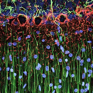
Background imageHistology Collection: Cerebellum tissue, light micrograph

Cerebellum tissue, light micrograph
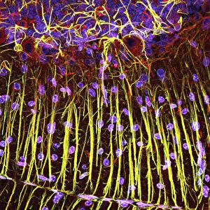
Cerebellum tissue. Confocal light micrograph of a section through the cerebellum of the brain. Purkinje cells, a type of neuron (nerve cell), are red

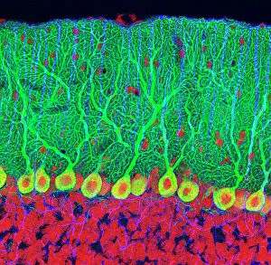
Background imageHistology Collection: Purkinje nerve cells in the cerebellum

Purkinje nerve cells in the cerebellum
Purkinje cells in the cerebellum. Fluorescent light micrograph of Purkinje cells (green) in the cerebellum of the brain. Purkinje nerve cells have a flask-like body from which numerous highly

Background imageHistology Collection: Hippocampus brain tissue

Hippocampus brain tissue
Hippocampus tissue. Light micrograph of a sagittal (side view) section through the hippocampus of the brain showing the nerve cells within it

Background imageHistology Collection: Cell types in the mammalian cerebellum: drawing, 1894, by the Spanish histologist Santiago Ramon y

Cell types in the mammalian cerebellum: drawing, 1894, by the Spanish histologist Santiago Ramon y Cajal (1852-1934)
NERVE CELLS, 1894. Cell types in the mammalian cerebellum: drawing, 1894, by the Spanish histologist Santiago Ramon y Cajal (1852-1934)

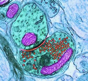
Background imageHistology Collection: Synapse nerve junction, TEM

Synapse nerve junction, TEM
Synapse. Coloured transmission electron micrograph (TEM) of a synapse, a junction between two nerve cells, in the brain. At a synapse an electrical signal is transmitted from one cell to the next in

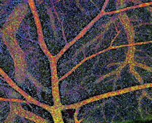
Background imageHistology Collection: Brain tissue blood supply

Brain tissue blood supply. Light micrograph of a section through cortex tissue from a brain, showing the blood vessels (branching) that supply it

Background imageHistology Collection: Kidney tubules in section

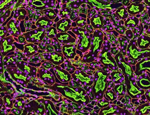
Kidney tubules in section
Kidney tubules. Fluorescent light micrograph of a section through kidney tissue showing numerous tubules (black/green). The tubules are seen in cross-section

Background imageHistology Collection: Human brain microscope slides

Human brain microscope slides. Microscope slides of stained sections of human brain. These slides are from the Harvard Brain and Tissue Resource Centre, USA

Background imageHistology Collection: Rough endoplasmic reticulum, TEM

Rough endoplasmic reticulum, TEM
Rough endoplasmic reticulum, coloured transmission electron micrograph (TEM). This section shows the rough endoplasmic reticulum (ER, folds, centre), a membranous structure that occurs in cells

Background imageHistology Collection: Glial stem cell culture, light micrograph

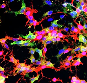
Glial stem cell culture, light micrograph
Glial stem cell culture. Fluorescent light micrograph of glial stem cells producing the protein NG2 (red) as they mature. These stem cells can differentiate into several types of glial cells

Background imageHistology Collection: Cerebral cortex nerve cells

Cerebral cortex nerve cells. Confocal light micrograph of neurons (nerve cells, red) and glial cells (support cells, gold) from the cerebral cortex

Background imageHistology Collection: Lactating breast tissue, light micrograph

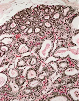
Lactating breast tissue, light micrograph
Lactating breast tissue. Light micrograph of a section through healthy breast (mammary gland) tissue during lactation. Glandular tissue in the breast has proliferated to produce spaces (alveoli)

Background imageHistology Collection: Purkinje nerve cells in the cerebellum

Purkinje nerve cells in the cerebellum
Purkinje cells in the cerebellum. Fluorescent light micrograph of Purkinje cells (green) in the cerebellum of the brain. Purkinje nerve cells have a flask-like body from which numerous highly

Background imageHistology Collection: Cerebellum structure, light micrograph

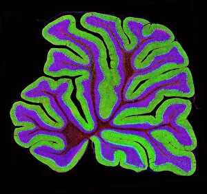
Cerebellum structure, light micrograph
Cerebellum structure. Fluorescent light micrograph of a section through the cerebellum of the brain. The cerebellum comprises three main layers

Background imageHistology Collection: Alzheimers brain

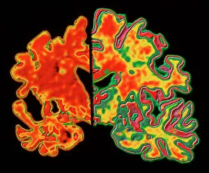
Alzheimers brain. Computer processed vertical (coronal) slice through the brain of an Alzheimer patient (at left) compared with a normal brain (at right)

Background imageHistology Collection: Pine pollen grains, light micrograph

Pine pollen grains, light micrograph
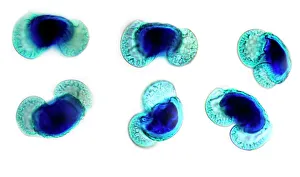
Pine pollen grains. Light micrograph of a section through winged pollen grains (microspores) from a pine (Pinus sp.) tree

Background imageHistology Collection: Pine stem, light micrograph

Pine stem, light micrograph
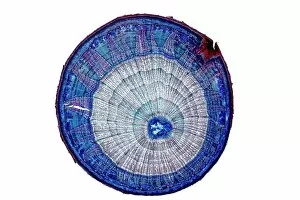
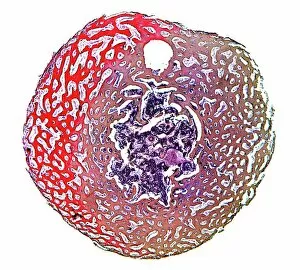
Pine stem. Light micrograph of a section through the stem of a pine (Pinus sp.) tree, showing xylem tissue. The xylem is made up of tracheid cells (light pink)

Background imageHistology Collection: Lime tree stem, light micrograph

Lime tree stem, light micrograph
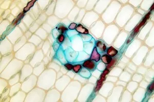
Lime tree stem. Light micrograph of a section through the stem of a lime tree (Tilia europaea). The outer epidermis has been shed and replaced by a layer of cork (dark red)

Background imageHistology Collection: Neural stem cell culture

Neural stem cell culture. Fluorescent light micrograph of a group of neural stem cells (neurosphere) in culture. Neural stem cells are able to differentiate into neurons (nerve cells)

Background imageHistology Collection: Compact bone, light micrograph

Compact bone, light micrograph
Compact bone. Polarised light micrograph of a transverse section through compact bone tissue, showing Haversian canals (circular regions)

Background imageHistology Collection: Fat cells, TEM

Fat cells, TEM
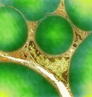
Fat cells. Coloured transmission electron micrograph (TEM) of a section through fat (adipose) tissue, consisting of fat-storing cells (adipocytes, green) supported by connective tissue

Background imageHistology Collection: Motor neurons, light micrograph

Motor neurons, light micrograph. Motor neurons are responsible for passing information around the central nervous system (CNS) and from the CNS to the rest of the body

Background imageHistology Collection: Cerebellum tissue, light micrograph

Cerebellum tissue, light micrograph
Cerebellum tissue. Confocal light micrograph of a section through the cerebellum of the brain showing two types of glial cells (support cells); astrocytes (star-shaped)

Background imageHistology Collection: Cerebellum tissue, light micrograph

Cerebellum tissue, light micrograph
Cerebellum tissue. Confocal light micrograph of a section through the cerebellum of the brain. Purkinje cells, a type of neuron (nerve cell), are red

Background imageHistology Collection: Plasma cells, TEM

Plasma cells, TEM
Plasma cells. Coloured transmission electron micrograph (TEM) of plasma cells. Plasma cells are mature B lymphocytes (white blood cells) that produce and secrete antibodies during an immune response

Background imageHistology Collection: Nerve cell, TEM

Nerve cell, TEM
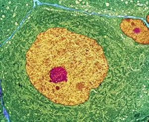
Nerve cell. Coloured transmission electron micrograph (TEM) of a nerve cell body in cross- section. The cell has a large nucleus (yellow) and inner nucleolus (red)

Background imageHistology Collection: Cystic fibrosis

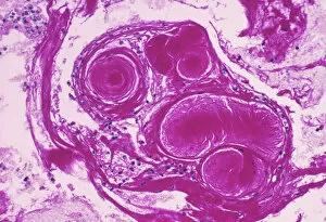
Cystic fibrosis. Light micrograph of a section through a bronchus (airway) of the lungs in a case of cystic fibrosis (mucoviscidosis)

Background imageHistology Collection: Islet of Langerhans, light micrograph

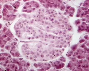
Islet of Langerhans, light micrograph
Islet of Langerhans. Light micrograph of a section through an islet of Langerhans (centre) in the pancreas. This structure is composed of clumps of secretory cells

Background imageHistology Collection: Collagen fibres

Collagen fibres. Coloured transmission electron micrograph (TEM) of a section through healthy collagen fibres from human skin

Background imageHistology Collection: Ovarian follicle, light micrograph C016 / 0519

Ovarian follicle, light micrograph C016 / 0519
Ovarian follicle. Light micrograph of a section through a secondary ovarian follicle showing signs of maturation into the antral follicle class

Background imageHistology Collection: Eye muscle, TEM C014 / 1468

Eye muscle, TEM C014 / 1468
Eye muscle. Transmission electron micrograph (TEM) of a section through a striated muscle cell from the ciliary muscle of a human eye

Background imageHistology Collection: Purkinje nerve cell, TEM C014 / 0583

Purkinje nerve cell, TEM C014 / 0583
Purkinje nerve cell. Transmission electron micrograph (TEM) of a purkinje nerve cell (green) from the cerebellum of the brain, showing the cell body (centre) and its primary dendrite (cell process)

Background imageHistology Collection: Skin layers, SEM

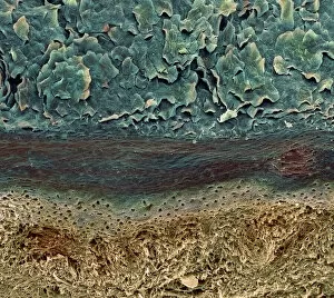
Skin layers, SEM

Background imageHistology Collection: Pancreatic islet of Langerhans

Pancreatic islet of Langerhans
Islet of Langerhans. Light micrograph of a section through an islet of Langerhans (pale, centre) in pancreas tissue. This clump of secretory cells forms part of the endocrine system of the body

Background imageHistology Collection: Blood vessel

Blood vessel. Coloured scanning electron micrograph (SEM) of a section through a blood vessel in the skin. The vessel contains red blood cells (erythrocytes, red). Magnification unknown

Background imageHistology Collection: Nerve synapse, TEM

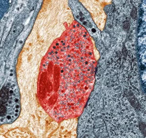
Nerve synapse, TEM
Nerve synapse. Coloured transmission electron micrograph (TEM) of the neuron (nerve) terminal at a synapse in the diaphragm

Background imageHistology Collection: Nerve cell, SEM

Nerve cell, SEM
Nerve cell. Coloured scanning electron micrograph (SEM) of a neuron (nerve cell). The cell body is the central structure with neurites (long and thin structures) radiating outwards from it

Background imageHistology Collection: Osteoporotic bone

Osteoporotic bone

Background imageHistology Collection: Intestinal microvilli, TEM

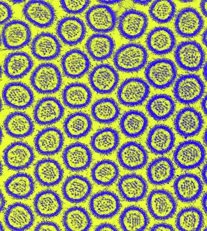
Intestinal microvilli, TEM
Intestinal microvilli, coloured transmission electron micrograph (TEM). The microvilli are finger-like projections (seen here in circular cross-section)

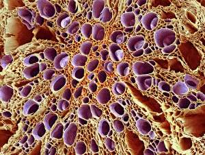
Background imageHistology Collection: Xylem tissue, SEM

Xylem tissue, SEM
Xylem tissue. Coloured scanning electron micrograph (SEM) of a section through xylem tissue from a dicotyledon rootlet. Xylem vessels (purple)

Background imageHistology Collection: Spongy bone, light micrograph

Spongy bone, light micrograph
Spongy bone. Light micrograph of a section through stained and decalcified human spongy bone. This bone type is also called cancellous bone

Background imageHistology Collection: Heart muscle, confocal light micrograph

Heart muscle, confocal light micrograph
Heart muscle. Confocal light micrograph of a section through cardiac (heart) muscle. Cardiac muscle consists of branching elongated muscle cells

Background imageHistology Collection: Histology of flowering plant (colour litho)

Histology of flowering plant (colour litho)
5200665 Histology of flowering plant (colour litho) by English School, (19th century); Private Collection; (add.info.: Histology of flowering plant)

Background imageHistology Collection: Charles-Francois Brisseau de Mirbel (engraving)

Charles-Francois Brisseau de Mirbel (engraving)
815844 Charles-Francois Brisseau de Mirbel (engraving) by French School, (19th century); Private Collection; (add.info.: Charles-Francois Brisseau de Mirbel (1776-18)

Background imageHistology Collection: Marie Francois Xavier Bichat, French physiologist and anatomist (chromolitho)

Marie Francois Xavier Bichat, French physiologist and anatomist (chromolitho)
1094403 Marie Francois Xavier Bichat, French physiologist and anatomist (chromolitho) by French School, (19th century); Private Collection; (add.info.: Marie Francois Xavier Bichat (1771-1802)

Background imageHistology Collection: SCHWANN, Theodor (1810 - 1882). German physiologist

SCHWANN, Theodor (1810 - 1882). German physiologist, anatomist and naturalist. He developed the cell theory. Oil

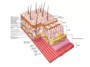
Background imageHistology Collection: Anatomy of the human skin

Anatomy of the human skin

Background imageHistology Collection: Medical ilustration of a pilonidal cyst near the natal cleft of the buttocks

Medical ilustration of a pilonidal cyst near the natal cleft of the buttocks


All Professionally Made to Order for Quick Shipping

Histology, the study of tissues at a microscopic level, unveils the intricate beauty and complexity of our body's structures. Through techniques like light micrography and transmission electron microscopy (TEM), scientists have been able to explore various tissues and unravel their secrets. One such tissue is the cerebellum, which plays a crucial role in coordinating movement and balance. By examining cerebellum tissue under a light microscope, we can observe its distinct layers and cell types. The synapse nerve junctions captured through TEM reveal the precise connections between neurons that allow for seamless communication. In 1894, Spanish histologist Santiago Ramon y Cajal created an exquisite drawing showcasing different cell types within the mammalian cerebellum. His meticulous work laid the foundation for understanding neural networks. Moving beyond just one region of the brain, histologists also delve into other fascinating areas like the hippocampus. Microscopic examination of hippocampus brain tissue provides insights into memory formation and spatial navigation. Purkinje nerve cells found within the cerebellum are particularly captivating under scrutiny. Their elaborate branching patterns give rise to their unique appearance when observed through a microscope slide. Histological studies extend beyond neurological tissues; they encompass organs throughout our body systems as well. For instance, kidney tubules in section offer glimpses into renal function while highlighting their structural organization. The human brain itself holds countless mysteries waiting to be unraveled by histologists examining microscope slides containing delicate slices of this complex organ. These slides provide glimpses into both healthy brains and those affected by diseases like Alzheimer's - offering valuable insights into neurodegenerative disorders. Exploring deeper with TEM reveals cellular components such as rough endoplasmic reticulum - an organelle involved in protein synthesis - providing detailed views at nanoscale resolution. Histology not only focuses on neurons but also encompasses glial cells that support neuronal functions. Light micrographs capturing glial stem cell cultures showcase the potential for regeneration and repair within the nervous system.