Fraud Blocker Skip to main content

Microvilli Collection

Microvilli are tiny, finger-like projections found on the surface of various cells in our body

Background imageMicrovilli Collection: Gallbladder surface, SEM

Gallbladder surface, SEM
Gallbladder. Coloured scanning electron micrograph (SEM) of the surface of the gallbladder showing the numerous microvilli that cover the columnar epithelium

Background imageMicrovilli Collection: Coloured SEM of a white blood cell (lymphocyte)

Coloured SEM of a white blood cell (lymphocyte)
Lymphocyte blood cell. Coloured scanning electron micrograph (SEM) of a lymphocyte, a common type of white blood cell. Characteristic of this rounded cell are the long microvilli which project

Background imageMicrovilli Collection: Goblet cells

Goblet cells. Coloured transmission electron micrograph (TEM) of a section through goblet cells in the lining of the small intestine, part of the digestive tract. They are full of mucus (yellow)

Background imageMicrovilli Collection: Sickle cell anemia with red blood cells and white bood cells

Sickle cell anemia with red blood cells and white bood cells
Conceptual image of sickle cell anemia with red blood cells and white bood cells

Background imageMicrovilli Collection: Model of animal cell, including cell nucleus, golgi body, lysosomes, centrioles, mitochondria

Model of animal cell, including cell nucleus, golgi body, lysosomes, centrioles, mitochondria, endoplasmic reticulum, ribosomes, cytoplasm, vesicles, thin plasma membrane

Background imageMicrovilli Collection: Lymphocyte white blood cell, SEM C016 / 9415

Lymphocyte white blood cell, SEM C016 / 9415
Lymphocyte white blood cell. Coloured scanning electron micrograph (SEM) of a lymphocyte white blood cell. Lymphocytes are involved in the immune systems defence mechanisms, lymph system

Background imageMicrovilli Collection: Intestinal microvilli, SEM C016 / 9066

Intestinal microvilli, SEM C016 / 9066
Intestinal microvilli. Coloured scanning electron micrograph (SEM) of a section through the lining of the small intestine, showing the densely packed microvilli (hair-like, top)

Background imageMicrovilli Collection: Intestinal microvilli, SEM C016 / 9067

Intestinal microvilli, SEM C016 / 9067
Intestinal microvilli. Coloured scanning electron micrograph (SEM) of a section through the lining of the small intestine, showing the densely packed microvilli (hair-like, top)

Background imageMicrovilli Collection: Intestinal brush border, TEM

Intestinal brush border, TEM
Intestinal brush border. Transmission electron micrograph (TEM) of intestinal absorptive cells sectioned horizontally at their apex to show the surface microvilli (round)

Background imageMicrovilli Collection: Gallbladder surface, SEM C015 / 9613

Gallbladder surface, SEM C015 / 9613
Gallbladder surface, coloured scanning electron micrograph (SEM). The columnar epithelium of the surface is covered in microvilli (tiny hair-like projections)

Background imageMicrovilli Collection: Gallbladder surface, SEM C015 / 9611

Gallbladder surface, SEM C015 / 9611
Gallbladder surface, coloured scanning electron micrograph (SEM). At centre are three secretory glands (yellow). The columnar epithelium of the surface is covered in microvilli

Background imageMicrovilli Collection: Gallbladder surface, SEM C015 / 9612

Gallbladder surface, SEM C015 / 9612
Gallbladder surface, coloured scanning electron micrograph (SEM). The columnar epithelium of the surface is covered in microvilli (tiny hair-like projections)

Background imageMicrovilli Collection: Trachea lining, TEM C014 / 1469

Trachea lining, TEM C014 / 1469
Trachea lining. Transmission electron micrograph (TEM) of a longitudinal section through the lining of the trachea (windpipe), which links the larynx (voicebox) to the lungs

Background imageMicrovilli Collection: Mouth cancer cell, SEM C014 / 1460

Mouth cancer cell, SEM C014 / 1460
Mouth cancer cell. Coloured scanning electron micrograph (SEM) of a squamous cell carcinoma (cancer) cell from a human mouth

Background imageMicrovilli Collection: Intestinal microvilli, SEM C014 / 1452

Intestinal microvilli, SEM C014 / 1452
Intestinal microvilli. Coloured transmission electron micrograph (SEM) of a transverse section through microvilli, showing their interiors

Background imageMicrovilli Collection: Mouth cancer cell, SEM C014 / 1459

Mouth cancer cell, SEM C014 / 1459
Mouth cancer cell. Coloured scanning electron micrograph (SEM) of a squamous cell carcinoma (cancer) cell from a human mouth

Background imageMicrovilli Collection: Dividing cancer cell, SEM C014 / 0362

Dividing cancer cell, SEM C014 / 0362
Dividing cancer cell. Coloured scanning electron micrograph (SEM) of a colorectal cancer cell undergoing mitosis (nuclear division) and splitting into two daughter cells (left and right)

Background imageMicrovilli Collection: Intestinal microvilli, SEM C014 / 1451

Intestinal microvilli, SEM C014 / 1451
Intestinal microvilli. Coloured transmission electron micrograph (SEM) of a transverse section through microvilli, showing their interiors

Background imageMicrovilli Collection: Trachea lining, TEM C014 / 1471

Trachea lining, TEM C014 / 1471
Trachea lining. Transmission electron micrograph (TEM) of a transverse section through the lining of the trachea (windpipe), which links the larynx (voicebox) to the lungs

Background imageMicrovilli Collection: Intestinal microvilli, TEM C014 / 1454

Intestinal microvilli, TEM C014 / 1454
Intestinal microvilli. Transmission electron micrograph (TEM) of a section through an epithelial cell from a human small intestine, showing the densely packed microvilli

Background imageMicrovilli Collection: Intestinal microvilli, TEM C014 / 1453

Intestinal microvilli, TEM C014 / 1453
Intestinal microvilli. Transmission electron micrograph (TEM) of a section through an epithelial cell from a human small intestine, showing the densely packed microvilli

Background imageMicrovilli Collection: Trachea lining, TEM C014 / 1470

Trachea lining, TEM C014 / 1470
Trachea lining. Transmission electron micrograph (TEM) of a longitudinal section through the lining of the trachea (windpipe), which links the larynx (voicebox) to the lungs

Background imageMicrovilli Collection: Trachea lining, TEM C014 / 1472

Trachea lining, TEM C014 / 1472
Trachea lining. Transmission electron micrograph (TEM) of a transverse section through the lining of the trachea (windpipe), which links the larynx (voicebox) to the lungs

Background imageMicrovilli Collection: Mouth cancer cells, SEM

Mouth cancer cells, SEM
Mouth cancer cells. Coloured scanning electron micrograph (SEM) of squamous cell carcinoma (cancer) cells from a human mouth

Background imageMicrovilli Collection: Mouth cancer cell dividing, SEM

Mouth cancer cell dividing, SEM
Mouth cancer dividing. Coloured scanning electron micrograph (SEM) of a squamous cell carcinoma (cancer) cell from a human mouth undergoing mitosis (nuclear division)

Background imageMicrovilli Collection: Mouth cancer cell, SEM

Mouth cancer cell, SEM
Mouth cancer cell. Coloured scanning electron micrograph (SEM) showing the surface of a squamous cell carcinoma (cancer) cell from a human mouth

Background imageMicrovilli Collection: Small bowel epithelium, TEM

Small bowel epithelium, TEM
Small bowel epithelium. Transmission electron micrograph (TEM) of a section through the epithelial lining of a villus in the small bowel, showing the enterocyte cells

Background imageMicrovilli Collection: Dividing cancer cell, SEM C014 / 0361

Dividing cancer cell, SEM C014 / 0361
Dividing cancer cell. Coloured scanning electron micrograph (SEM) of a colorectal cancer cell undergoing mitosis (nuclear division) and splitting into two daughter cells (left and right)

Background imageMicrovilli Collection: Dividing brain cancer cells, SEM C014 / 0354

Dividing brain cancer cells, SEM C014 / 0354
Dividing brain cancer cells. Coloured scanning electron micrograph (SEM) of a cancerous astrocyte brain cell that has just undergone cytokinesis (cell division)

Background imageMicrovilli Collection: Cancer cell, SEM C014 / 0628

Cancer cell, SEM C014 / 0628
Cancer cell. Coloured scanning electron micrograph (SEM) of a carcinoma cancer cell (pink) migrating (metastasising) along a layer of normal epithelial cells (purple)

Background imageMicrovilli Collection: Cancer cell, SEM C014 / 0627

Cancer cell, SEM C014 / 0627
Cancer cell. Coloured scanning electron micrograph (SEM) of a carcinoma cancer cell (yellow) migrating (metastasising) along a layer of normal epithelial cells (brown)

Background imageMicrovilli Collection: TEM of Giardia lamblia

TEM of Giardia lamblia
Coloured Transmission Electron Micrograph of Giardia lamblia (orange), a parasitic flagellate protozoan which causes the disease lambliasis (giardiasis)

Background imageMicrovilli Collection: Kidney tubule, TEM

Kidney tubule, TEM
Kidney tubule. Coloured transmission electron micrograph (TEM) of a section through a proximal convoluted tubule in the kidney

Background imageMicrovilli Collection: Duodenal microvilli

Duodenal microvilli
Microvilli in duodenum. Coloured transmission electron micrograph (TEM) of a section through the human duodenum, showing microvilli on the surface epithelium (lining)

Background imageMicrovilli Collection: Coloured TEM of microvilli of intestinal cell

Coloured TEM of microvilli of intestinal cell

Background imageMicrovilli Collection: Fallopian tube

Fallopian tube. Coloured scanning electron micrograph (SEM) of the epithelium of a fallopian tube (oviduct), one of two muscular tubes connecting the ovary to the uterus (womb)

Background imageMicrovilli Collection: Small intestine villus, SEM

Small intestine villus, SEM
Small intestine villus. Coloured scanning electron micrograph (SEM) of a freeze fracture section through a villus from the mucosal lining of the small intestine

Background imageMicrovilli Collection: Coloured SEM of fallopian tube cilia & microvilli

Coloured SEM of fallopian tube cilia & microvilli
Fallopian tube cells. Coloured high resolution scanning electron micrograph of cilia and micro- villi on the surface of the fallopian tube epithelium

Background imageMicrovilli Collection: Trachea lining, TEM

Trachea lining, TEM
Trachea lining. Coloured transmission electron micrograph (TEM) of a transverse section through the lining of the trachea (windpipe). The trachea links the larynx to the lungs


All Professionally Made to Order for Quick Shipping

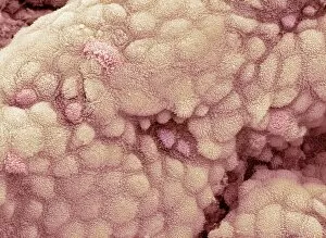
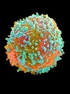
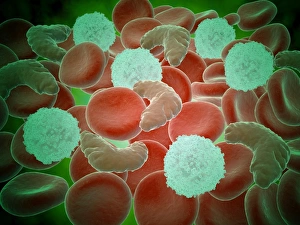
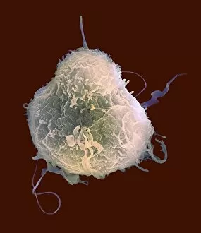
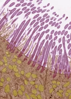
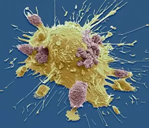
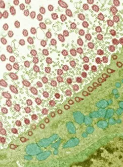
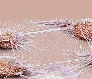
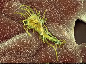
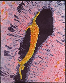
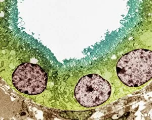
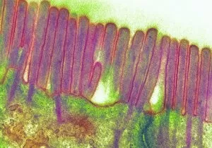
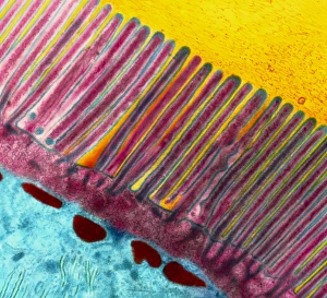
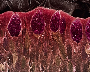
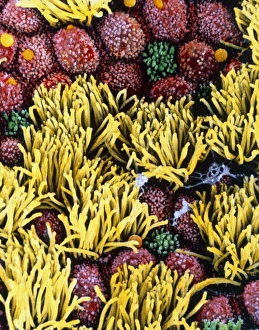
Microvilli are tiny, finger-like projections found on the surface of various cells in our body. When observed under a scanning electron microscope (SEM), these microvilli appear as delicate structures covering the gallbladder surface, enhancing its absorptive capacity. Similarly, a colored SEM image reveals their presence on white blood cells known as lymphocytes, suggesting their role in immune responses. Interestingly, it also play a crucial role in intestinal protozoan parasites' attachment and invasion into host cells. This is evident from transmission electron microscopy (TEM) images showcasing these parasites exploiting microvilli to establish infections. In sickle cell anemia patients, microscopic examination showcases red and white blood cells affected by this genetic disorder. Amongst the components visible in the model of an animal cell are microvilli protruding from the top surface. These structures aid in increasing the cell's surface area for absorption and secretion processes. The significance extends beyond normal cellular functions; they can be observed even during pathological conditions like vaginal cancer or Chlamydia infection. Scanning electron microscopy captures detailed images of cancerous vaginal cells with distinct microvilli patterns associated with disease progression. Furthermore, SEM images provide insights into foetal skin development by revealing intricate arrangements of developing tissues along with prominent microvillar formations that contribute to nutrient exchange between mother and fetus.